

The Diagnosis and Treatment of Hemoptysis
- PMID: 28625277
- PMCID: PMC5478790
- DOI: 10.3238/arztebl.2017.0371
The Diagnosis and Treatment of Hemoptysis
Abstract
Background: Hemoptysis, i.e., the expectoration of blood from the lower airways, has an annual incidence of approximately 0.1% in ambulatory patients and 0.2% in inpatients. It is a potentially life-threatening medical emergency and carries a high mortality.
Methods: This review article is based on pertinent publications retrieved by a selective search in PubMed.
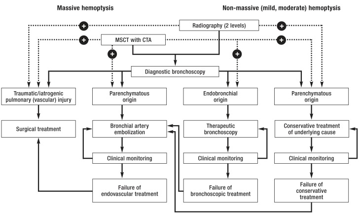
Results: Hemoptysis can be a sign of many different diseases. Its cause remains unknown in about half of all cases. Its more common recognized causes include infectious and inflammatory airway diseases (25.8%) and cancer (17.4%). Mild hemoptysis is self-limited in 90% of cases; massive hemoptysis carries a worse prognosis. In patients whose life is threatened by massive hemoptysis, adequate oxygenation must be achieved through the administration of oxygen, positioning of the patient with the bleeding side down (if known), and temporary intubation if necessary. A thorough diagnostic evaluation is needed to identify the underlying pathology, site of bleeding, and vascular anatomy, so that the appropriate treatment can be planned. The evaluation should include conventional chest x-rays in two planes, contrastenhanced multislice computerized tomography, and bronchoscopy. Hemostasis can be achieved at bronchoscopically accessible bleeding sites with interventionalbronchoscopic local treatment. Bronchial artery embolization is the first line of treatment for hemorrhage from the pulmonary periphery; it is performed to treat massive or recurrent hemoptysis or as a presurgical measure and provides successful hemostasis in 75-98% of cases. Surgery is indicated if bronchial artery embolization alone is not successful, or for special indications (traumatic or iatrogenic pulmonary/vascular injury, refractory asper gilloma).
Conclusion: The successful treatment of hemoptysis requires thorough diagnostic evaluation and close interdisciplinary collaboration among pulmonologists, radiologists, and thoracic surgeons.
Figures
Comment in
-
Particularities of Goodpasture Syndrome.Dtsch Arztebl Int. 2017 Sep 29;114(39):662. doi: 10.3238/arztebl.2017.0662a. Dtsch Arztebl Int. 2017. PMID: 29034872 Free PMC article. No abstract available.
References
-
- Jeudy J, Khan AR, Mohammed TL, et al. ACR appropriateness criteria hemoptysis. J Thorac Imaging. 2010;25:67–69. - PubMed
-
- Costabel U, Kroegel C. Klinische Pneumologie: Das Referenzwerk für Klinik und Praxis. Thieme. 2013
-
- Earwood JS, Thompson TD. Hemoptysis: evaluation and management. Am Fam Physician. 2015;91:243–249. - PubMed
-
- Abdulmalak C, Cottenet J, Beltramo G, et al. Haemoptysis in adults: a 5-year study using the French nationwide hospital administrative database. Eur Respir J. 2015;46:503–511. - PubMed
Publication types
MeSH terms
LinkOut - more resources
Full Text Sources
Other Literature Sources