. 2017 Jul 5;95(1):180-194.e5.
doi: 10.1016/j.neuron.2017.05.019. Epub 2017 Jun 15.

A Corticothalamic Circuit for Dynamic Switching between Feature Detection and Discrimination

Affiliations

A Corticothalamic Circuit for Dynamic Switching between Feature Detection and Discrimination

Wei Guo et al. Neuron. .

Abstract

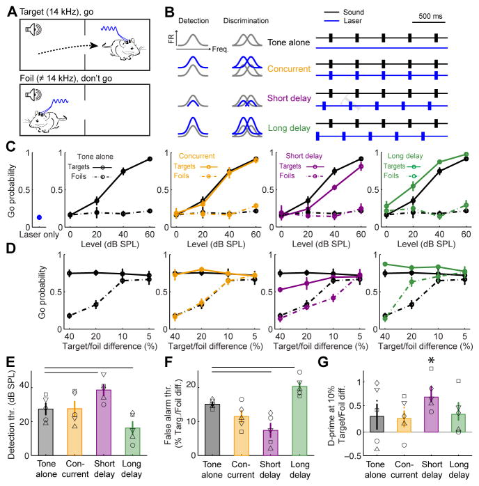
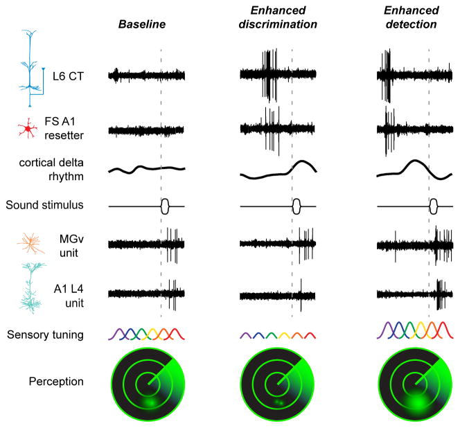
Sensory processing must be sensitive enough to encode faint signals near the noise floor but selective enough to differentiate between similar stimuli. Here we describe a layer 6 corticothalamic (L6 CT) circuit in the mouse auditory forebrain that alternately biases sound processing toward hypersensitivity and improved behavioral sound detection or dampened excitability and enhanced sound discrimination. Optogenetic activation of L6 CT neurons could increase or decrease the gain and tuning precision in the thalamus and all layers of the cortical column, depending on the timing between L6 CT activation and sensory stimulation. The direction of neural and perceptual modulation - enhanced detection at the expense of discrimination or vice versa - arose from the interaction of L6 CT neurons and subnetworks of fast-spiking inhibitory neurons that reset the phase of low-frequency cortical rhythms. These findings suggest that L6 CT neurons contribute to the resolution of the competing demands of detection and discrimination.

Keywords: auditory cortex; auditory thalamus; delta rhythm; layer 6; medial geniculate body; modulation; oscillation; phase reset; plasticity; theta rhythm.


Figures

Figure 1
Figure 1. A transgenic strategy to selectively target layer 6 auditory corticothalamic neurons (L6 CT)
(A) Ntsr1-Cre mice were crossed with a Cre-dependent tdTomato reporter line. Fluorescent microspheres injected into the medial geniculate body (MGB) were retrogradely transported to the cell bodies of CT neurons in the auditory cortex. (B) After allowing the thalamic beads 7 days for retrograde transport, coronal sections of auditory cortex were immunolabeled for NeuN, a ubiquitous neural marker. As expected, CT cell bodies identified with green beads were occasionally found in L5 (white arrow) but were concentrated in L6, where Ntsr1+ cell bodies (red) were abundant. Scale bar = 100 μm. (C) Magnification of area designated by white square in (B). Many L6 neurons (blue channel, top left) are Ntsr1+ (red channel, top right) and project to a MGB region near the retrobead injection zone (green channel, bottom left). L6 neurons that do not project to the MGB and do not express Ntsr1-Cre (gray arrowhead, bottom right) are interspersed among Ntsr1+ L6 CT neurons (white arrowhead). Scale bar = 10 μm. (D) We quantified the percentage of L6 neurons that were CT and/or Ntsr1+ in 824 L6 neurons from 8 hemispheres of 4 mice. (E–F) Summary histograms for the percentage of neurons that were positive or negative for beads (E) or Ntsr1 (F). Approximately 35% of L6 neurons do not express Ntsr1 and do not project to the MGB retrobead injection zone (left bars). By contrast, 97% of L6 Ntsr1+ neurons project to the MGB, confirming that Ntsr1-Cre selectively targets L6 CT neurons in the auditory cortex.
Figure 2
Figure 2. Optogenetic activation of L6 CT neurons in A1 induces alternating periods of spike enhancement and suppression
(A) ChR2 was expressed in L6 CT neurons by injecting a Cre-dependent viral construct into A1 of Ntsr1-Cre mice. The fluorescent mCherry reporter is visible in L6 cell bodies, dense bands of neuropil in L4 and in MGB axons. (B) Schematic of columnar recording during L6 CT activation. (C) Sound-evoked (left) and laser-evoked (right) laminar profiles of current source density (CSD) amplitude from a single A1 penetration in an awake mouse. Multiunit activity (MUA) at each location is represented by the superimposed white peristimulus time histograms (PSTH, scale bar = 100 spikes/s). White arrow indicates a brief, early sound-evoked current sink used to identify L4. (D) PSTHs represent the mean MUA in each layer. Laser power at the tip of the optic fiber ranges from 5–50 mW. Error bars represent 1 SEM. (E) The change in laser-evoked firing rates relative to baseline activity increase monotonically during the onset and sustained periods (ANOVA, F > 10, p < 1 x 10⁻⁶ for each, orange and gray arrows, respectively), decrease immediately following laser offset (F=22.99, p < 1 x 10⁻⁶, purple arrow) and increase again 100ms later (F=4.01, p < 5 x 10⁻⁵, green arrow), particularly in L2/3, L4 and L5. Error bars represent 1 SEM. A detailed breakdown of all statistical tests can be found in Supplemental Table 1.
Figure 3
Figure 3. Activating L6 CT neurons induces a tri-phasic modulation of sound-evoked A1 responses
(A) Pure tone pips were presented to the left ear while recording spiking activity from the contralateral A1 with and without optogenetic activation of L6 CT neurons. Frequency response functions are illustrated with a Gaussian fit. (B) Tones were presented without L6 CT activation (50% of trials, gray) or with L6 CT activation, where the delay between the onset of tone bursts and laser pulses varied from 0 – 800 ms. (C) L6 CT activation imposed diverse forms of modulation on a representative L4 recording site. (D–E) The scaling and shifting components were computed in the same example L4 site (D) and a representative L6 site (E) by regressing the mean normalized tone-evoked firing rates measured during the tone-alone trials against the firing rates recorded during the three epochs surrounding L6 CT activation. (F–G) Mean (±1 SEM) shifting and scaling modulation were computed for each multiunit site for recordings made with a 400 ms laser pulse at 20 mW (F) or a 50 ms laser pulse set to a minimally effective laser power (G). Inset: Laser-induced tuning modulation was not observed from a separate cohort of mice injected with a control virus (Mixed design ANOVA, main effect for shifting modulation: F(2,20) = 0.24, p = 0.79; main effect for scaling modulation: F(2,20) = 2.17, p = 0.14). Statistically significant shifting and scaling modulation for all permutations of laser duration, response period and layer were determined with one-sample t-tests against a population mean of 0 (shifting modulation) or 1.0 (scaling modulation). Looking across both laser durations, we observed significant additive changes during L6 CT activation in L2/3, L5 and L6 (p < 0.05 for each) and significant divisive gain in L4 and L5 (p < 0.05 for each). In the short-delay period following L6 CT deactivation, we observed significant subtractive changes in all layers and significant divisive gain in all but L6 (p < 0.005 for each). In the long-delay period following L6 CT deactivation, we observed significant additive and multiplicative changes for L2/3, L4 and L5 (p < 0.05 for each) but no significant change in L6 tuning (p > 0.1 for each). (H) The mixture of shifting and scaling modulation created sharper frequency tuning during the short-delay period but wider frequency tuning in the long-delay period. Tuning changes in octaves (oct.) are estimated from the change in width at half-height between the tone-alone and tone + L6 CT activation for the 400 ms laser (top) and 50 ms laser (bottom) conditions. Asterisks indicate p < 0.05 with a one-sample t-test relative to a population mean of 0.
Figure 4
Figure 4. L6 CT activation can bias sound perception towards enhanced detection at the expense of discrimination, or vice versa
(A) Mice were trained in an auditory avoidance task that required them to cross from one side of a shuttle box shortly following the presentation of 14 kHz tone bursts (target) but not tones of other frequencies (foils). Mice expressed ChR2 in L6 CT neurons in left and right auditory cortex and were implanted with bilateral optic fiber assemblies. (B) Schematic of A1 tuning modulation and design of behavioral optogenetics experiment. The distinct types of receptive field modulation following L6 CT deactivation were predicted to have dissociable effects on tone detection and discrimination behaviors. (C) Probability of a “Go” (i.e., crossing) response for target tones, foil tones, the laser stimulus alone and the three combined tone + laser test conditions as a function of sound level. Compared to tone-alone trials, target detection is impaired in the short-delay configuration but enhanced in the long-delay configuration (ANOVA, main effects for delay, F = 10.44, p < 0.005 for short and long conditions). (D) Probability of a Go response as a function of frequency separation between the target tone and the foil tone at a fixed sound level (40 dB SPL). Discrimination is enhanced for difficult conditions (10%) in the short delay condition but is reduced in easy conditions (20%) in the long delay condition, ANOVA, main effect for delay condition, F = 14.3, p < 0.0005 for both short and long delays). (E) Mean (±1 SEM) target detection threshold (thr.), defined as the sound level associated with a 50% probability of making a Go response on target trials. (F) Mean (±1 SEM) false alarm threshold, defined as the frequency spacing associated with a 50% probability of making a Go response on foil trials. Horizontal lines in E and F represent p < 0.05 using a paired t-test between tone-alone and the corresponding tone + laser condition, after correcting for multiple comparisons. (G) Overall sensitivity, measured with the d’ statistic, was higher on short-delay trials, but the difference is not statistically significant after correcting for multiple comparisons (paired t-test, p = 0.16). However, the d’ statistic was significantly different than zero (no separation between the hit and false positive distributions) for the short-delay period, indicated by asterisk (one-sample t-test, p < 0.001, p > 0.1 for all other conditions).
Figure 5
Figure 5. Enhanced A1 responses at long delays following L6 CT activation can be attributed to short-term dynamics in thalamic sound processing
(A) Coronal sections showing mCherry expression in auditory L6 CT neurons and medial geniculate body (MGB) axon terminals (top) as well as a more rostral section showing the L6 CT axon bundle in the internal capsule (ic) and collaterals in the thalamic reticular nucleus (TRN, bottom). (B) Schematic of procedure for simultaneous recordings of the A1 column and either MGB or TRN in awake, head-fixed mice. The ventral, medial and dorsal subdivisions of the MGB are illustrated (v, m and d, respectively). Parameters for L6 CT activation laser were a 50 ms duration laser pulse, 5 mW above threshold. (C) Frequency response areas (FRAs) from simultaneously recorded A1/MGB or A1/TRN sites. Recordings were topographically aligned such that frequency tuning was roughly matched between cortical and thalamic sites. (D) Iso-intensity frequency tuning functions from representative L4, MGB and TRN recording sites. The gray and blue functions correspond to the tone-alone and tone + laser conditions, respectively. Enhanced auditory responses are observed in both A1 and thalamus when tones and L6 CT activation are concurrent (orange). Divisive suppression is found in A1 shortly after L6 CT deactivation but not in either thalamic recording site (purple). Multiplicative enhancement is observed in A1 and MGB at long delays following L6 CT deactivation (green). TRN tuning is suppressed at this interval. (E) Mean (±1 SEM) tone-evoked firing rates normalized to the best frequency in the tone-alone condition (gray). Compared to tone-alone responses, firing rates were significantly elevated with concurrent L6 CT activation in A1, MGB and TRN units (paired t-test, p < 0.05); during the short delay period, A1 and TRN units showed significantly reduced firing rates (paired t-test, p < 0.05) while MGB units showed unchanged firing rates (p > 0.05); during the long delay period, A1 and MGB units showed significantly enhanced firing rates (p < 0.05) while TRN units showed significantly reduce firing rates (p < 0.05). (F) Mean (±1 SEM) shifting and scaling modulation computed for each multiunit site with the paired recording approach. Statistically significant shifting and scaling modulation for all permutations of laser duration, response period and brain structure was determined with one-sample t-tests against a population mean of 0 (shifting modulation) or 1.0 (scaling modulation). We observed significant additive gain during L6 CT activation in L2–5 and MGB and significant divisive gain in L2–5 (p < 0.05 for each). In the short-delay period following L6 CT deactivation, we observed significant subtractive and divisive gain only in L2–5 (p < 0.05 for both). In the long-delay period following L6 CT deactivation, we observed significant additive gain in MGB and L2–5, significant multiplicative gain in L2–5 and MGB, and significant subtractive and divisive gain in TRN (p < 0.05 for each).
Figure 6
Figure 6. L6 CT activation changes the frequency and resets the phase of local electric field oscillations in A1
(A) The raw L6 CT-evoked CSD signal recorded across the A1 column from a single trial in an awake mouse. Optogenetic activation of L6 CT neurons induces a high-frequency oscillation while the laser is on, followed by a few cycles of a low-frequency rhythm following L6 CT deactivation. Scale bar = 0.2 s and 5 mV/mm2. (B–C) Change in CSD frequency power spectrum during laser (blue) and 0–400 ms after the laser is turned off (black) relative to pre-laser baseline. B plots the full frequency range to highlight the high-gamma peak during laser activation, whereas C plots frequencies ≤ 30 Hz to emphasize the delta-theta power after laser offset. (D) Mean (±1 SEM) L2/3 unfiltered CSD amplitude for laser durations varying from 10–400 ms. The short- and long-delay periods following L6 CT deactivation are indicated by the purple and green arrows, respectively. (E) Phase histograms at the corresponding laser duration for the short- and long-delay period. L2/3 CSD phase is consistently near zero in the short-delay period and pi in the long-delay period. (F) Normalized spontaneous firing rate in each layer as a function of the spontaneous L2/3 delta-theta CSD phase (2–6 Hz). Spontaneous firing rate was modulated across L2/3 CSD phase for all layers (ANOVA, F > 3.6 and p < 0.001), with the lowest spike rate consistently occurring at the zero phase. (G) Tone-evoked frequency tuning functions at three phases of the spontaneously occurring L2/3 delta-theta CSD: zero, π and the average of the intermediate phases, ±π/2. Tuning shape was significantly modulated by phase in L4, L5 and L6, (ANOVA, F > 6.3, p < 0.005 for all), but not in L2/3 itself (F = 1.4, p = 0.28).
Figure 7
Figure 7. L6 CT neurons reset the phase of low-frequency cortical rhythms by driving a sub-type of cortical fast-spiking interneuron
(A) Schematic diagram depicting the analysis approach for spike-triggered CSD phase and amplitude. (B) Spike-triggered CSD amplitude from two exemplar A1 single units. Spontaneous spikes are associated with a clear pattern of alternating sinks and sources across the cortical column in some single units (a “resetter” unit, bottom), but not others (a “non-resetter” unit, top). (C) L2/3 delta-theta CSD phase histograms at discrete time bins before and after spontaneous spikes from the same two single units shown in (B). The L2/3 CSD vector strength represents the phase precision over time. (D) Spontaneous spike-triggered phase changes from 723 single units recorded from the thalamus and cortex of awake mice are sorted according to the change in L2/3 CSD vector strength. Resetter units (n = 184) were operationally defined as those that increased the post-spike L2/3 CSD vector strength by 0.05 or more. (E) Spike-triggered phase histograms from all resetter and 185 non-resetter units. Each trace is the mean phase trajectory from a single unit. Resetter units reset the phase of the L2/3 CSD to π at the time of the spike. The spike-triggered phase remains well organized at the short- and long-delay intervals following spontaneous resetter spikes (purple/pi and green/0 phase, respectively). (F) Histogram of resetter occurrence as a function of brain region and spike waveform. G) Mean (±1 SEM) latency between spontaneous spike occurrence and the time of CSD reset, operationally defined as the peak negativity in the L2/3 delta-theta CSD waveform. For MGBv resetter (MGBvr, n = 52) and cortical FSr units (n=31), spontaneously occurring spikes occured significantly earlier than the L2/3 CSD reset, suggesting that they could induce the reset (paired t-tests, p < 0.05 for both). By contrast, RSr unit spikes (n=78) occurred during or just after the CSD reset event (paired t-test, p = 0.1). (H) Cartoon illustrating the 4 cell types in the putative CSD reset circuit and the cross-correlation analysis approach. (I) Mean (±1 SEM) cross-correlogram of L6 CT unit spike trains with the other resetter unit types. (J) Mean (±1 SEM) probability that a spike in each resetter type will follow a laser-evoked L6 CT spike. Horizontal lines represent significant differences between FSr evoked spike probability and other unit types (unpaired t-test, p < 0.05 after correcting for multiple comparisons). (K) FSr-spike triggered L2/3 CSD amplitude for spike events occurring spontaneously, during auditory stimulation or during optogenetic activation of L6 CT units. Note similarity of FSr-triggered CSD change and laser-evoked CSD change in FIg. 6D. (L) Mean (±1 SEM) laser-evoked firing rate in L6 CT neurons, FSr neurons and FS neurons not associated with CSD reset (FSnr). (M) L6 CT spiking ceases immediately at laser offset whereas FSr neuron spiking remains significantly higher for at least 10ms (Wilcoxon rank-sum, p < 0.05).
Figure 8
Figure 8. Summary of findings supporting a contribution of L6 CT neurons to perceptual modes of heightened detection or discrimination
Unit recording traces are arranged to illustrate the main effects described in previous figures. Left column, in a baseline condition with minimal spiking activity in L6 CT and FS resetter neurons, the power of low-frequency CSD rhythms is weak and sound-evoked spiking in thalamic and cortical principal neurons is moderate. Intense firing of L6 CT neurons engages FS resetter interneurons that increase the power and reset the phase of low-frequency rhythms. Middle columns, at short delays following an intense volley of spikes in L6 CT and FS resetter neurons, the induced cortical delta-theta rhythm is at a positive, low-excitability phase and sound-evoked spikes are suppressed in A1, but in the MGBv. Right column, at longer delays following a volley of spikes in L6 CT and FS resetter neurons, the phase of the cortical delta-theta rhythm has rotated to a negative, high-excitability phase and sound-evoked spikes are greatly enhanced both in MGBv and A1. Modulating the excitability of cortical neurons has predictable effects on sensory tuning across ensembles of A1 neurons and behavioral measures of sound perception, illustrated as the resolution of two objects on a sonar display. Suppressed sound-evoked A1 activity shortly following the volley of spikes in the cortical reset network dampens excitability but reduces overlap between neighboring tuning functions, supporting enhanced discrimination of sound frequencies but reduced auditory sensitivity (middle column). Enhanced sound-evoked spiking scales up excitability and increases the overlap between neighboring tuning functions, resulting in enhanced sensitivity to sound at the expense of reduced frequency discriminability (right column).

References

    1. Bortone DS, Olsen SR, Scanziani M. Translaminar inhibitory cells recruited by layer 6 corticothalamic neurons suppress visual cortex. Neuron. 2014;82:474–485. - PMC - PubMed
    1. Bourassa J, Deschenes M. Corticothalamic projections from the primary visual cortex in rats: a single-fiber study using biocytin as an anterograde tracer. Eur J Neurosci. 1995;7:19–30. - PubMed
    1. Briggs F, Kiley CW, Callaway EM, Usrey WM. Morphological substrates for parallel streams of corticogeniculate feedback originating in both V1 and V2 of the macaque monkey. Neuron. 2016;90:388–399. - PMC - PubMed
    1. Buran BN, von Trapp G, Sanes DH. Behaviorally gated reduction of spontaneous discharge can improve detection thresholds in auditory cortex. J Neurosci. 2014;34:4076–4081. - PMC - PubMed
    1. Carracedo LM, Kjeldsen H, Cunnington L, Jenkins A, Schofield I, Cunningham MO, Davies CH, Traub RD, Whittington M. A neocortical delta rhythm facilitates reciprocal interlaminar interactions via nested theta rhythms. J Neurosci. 2013;33:10750–10761. - PMC - PubMed

STAR Methods References

    1. Anderson LA, Linden JF. Physiological differences between histologically defined subdivisions in the mouse auditory thalamus. Hear Res. 2011;274:48–60. - PMC - PubMed
    1. Hackett TA, Rinaldi Barkat T, O’Brien BMJ, Hensch TK, Polley DB. Linking topography to tonotopy in the mouse auditory thalamocortical circuit. J Neurosci. 2011;31:2983–2995. - PMC - PubMed
    1. Kaur S, Rose HJ, Lazar R, Liang K, Metherate R. Spectral integration in primary auditory cortex: Laminar processing of afferent input, in vivo and in vitro. Neuroscience. 2005;134:1033–1045. - PubMed