Review
. 2017 Jul;69(3):256-297.
doi: 10.1124/pr.116.013367.

The Diverse Roles of Arrestin Scaffolds in G Protein-Coupled Receptor Signaling

Affiliations
Review

The Diverse Roles of Arrestin Scaffolds in G Protein-Coupled Receptor Signaling

Yuri K Peterson et al. Pharmacol Rev. 2017 Jul.

Abstract

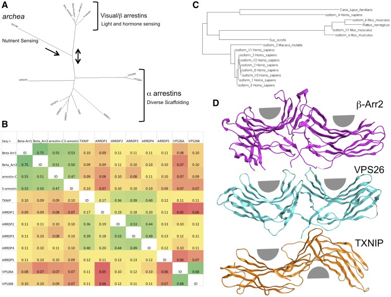
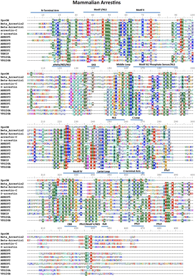
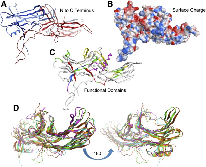
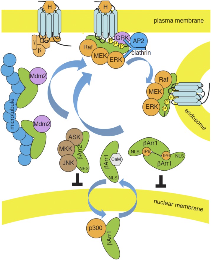
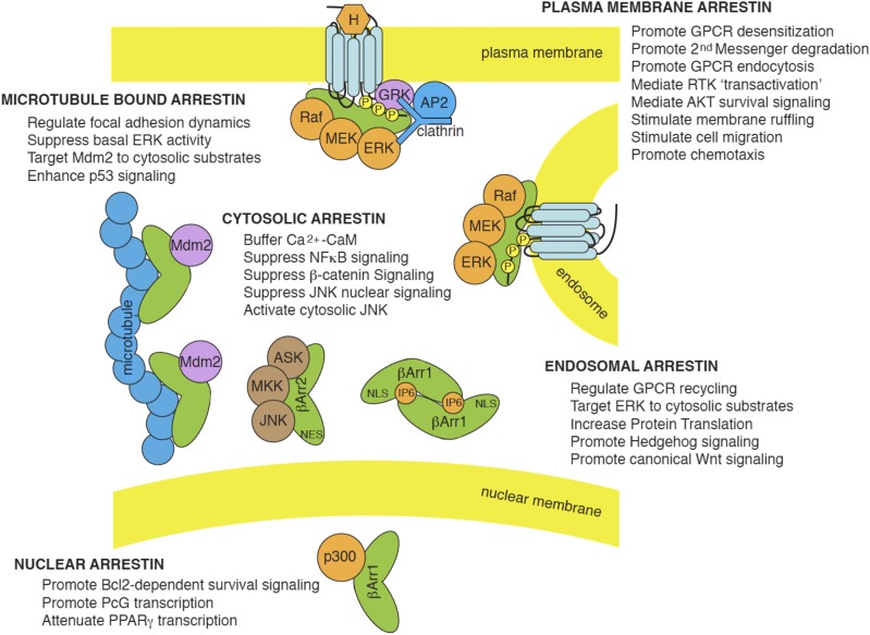
The visual/β-arrestins, a small family of proteins originally described for their role in the desensitization and intracellular trafficking of G protein-coupled receptors (GPCRs), have emerged as key regulators of multiple signaling pathways. Evolutionarily related to a larger group of regulatory scaffolds that share a common arrestin fold, the visual/β-arrestins acquired the capacity to detect and bind activated GPCRs on the plasma membrane, which enables them to control GPCR desensitization, internalization, and intracellular trafficking. By acting as scaffolds that bind key pathway intermediates, visual/β-arrestins both influence the tonic level of pathway activity in cells and, in some cases, serve as ligand-regulated scaffolds for GPCR-mediated signaling. Growing evidence supports the physiologic and pathophysiologic roles of arrestins and underscores their potential as therapeutic targets. Circumventing arrestin-dependent GPCR desensitization may alleviate the problem of tachyphylaxis to drugs that target GPCRs, and find application in the management of chronic pain, asthma, and psychiatric illness. As signaling scaffolds, arrestins are also central regulators of pathways controlling cell growth, migration, and survival, suggesting that manipulating their scaffolding functions may be beneficial in inflammatory diseases, fibrosis, and cancer. In this review we examine the structure-function relationships that enable arrestins to perform their diverse roles, addressing arrestin structure at the molecular level, the relationship between arrestin conformation and function, and sites of interaction between arrestins, GPCRs, and nonreceptor-binding partners. We conclude with a discussion of arrestins as therapeutic targets and the settings in which manipulating arrestin function might be of clinical benefit.


Figures

Fig. 1.
Fig. 1.
Evolutionary relationship of proteins sharing the common arrestin fold. (A) Radial phylogram showing the relation of all mammalian arrestins to the archaea arrestin-like molecule SpoOM. It also shows the relation to other mammalian arrestin family members, including the VPS and TXNIP proteins, which suggest that arrestins evolved from a mechanism to regulate environmental sensing toward more complicated signaling. NCBI BLAST was queried using the human β-arrestin2 full-length sequence. Psi-BLAST was used to search the RefSeq database. The sequence analysis was performed using Fitch phylogenetic tree analysis implemented in BioEdit 7.2.5 and visualized using Dendroscope 3.2.4. This graph was inspired from (Alvarez, 2008; Aubry et al., 2009). (B) Sequence identity matrix indicating the overall similarity of human arrestins. The analysis was performed using BLOSOM62 implemented in BioEdit 7.2.5. Cells were colored as a heat map using EXCEL according to percent similarity, with green being most similar and red being divergent. (C) Rectangular phylogram showing the relation of mammalian β-arrestin2 isoforms across several species. It demonstrates an expansion of diversity in humans and mice that may be indicative of the need to regulate complex hormone signaling and olfaction. The sequence analysis was performed using Fitch phylogenetic tree analysis implemented in BioEdit 7.2.5 and visualized using Dendroscope 3.2.4. (D) Structural comparison of β-arrestin, VPS26, and TXNIP showing different rotations between the amino and carboxy arrestin domains. VPS26 β-baskets are nearly symetirical and on the same plane, and β-arrestins show a compaction of the amino-terminal domain and a slight rotation between domains, whereas TXNIP has an almost 180° degree inversion of the relative position of the β-baskets. Structural PDB files (2WTR, 4P2A, and 4LL4) were superposed using only the amino-terminal domain to highlight the relative differences in the carboxy domain. Gray half circles were added to show the relative positions of the concave portion of the two β sandwiches per molecule. Analysis and visualization were performed using MOE 2014.09.
Fig. 2.
Fig. 2.
Amino acid sequence alignment of arrestins indicating extensions, insertions, deletions, and functional domains among the arrestin clade. Domains depicted include the N- and C- terminal arms, Motif I-IV, NES, three NLS, SH3 domain, four PPxY motifs, clathrin binding domain (CBD), and the middle, C, and Lariat loops. The analysis was performed using ClustalW multiple alignment analysis implemented in BioEdit 7.2.5. Amino acids are colored according to their chemical properties, and conserved consensus resides are colored filled.
Fig. 3.
Fig. 3.
Visual/β-arrestin topologic structural analysis showing the overall tertiary fold of arrestins, the charge distribution surface, and the major functional and interaction domains. (A) Ribbon diagram indicating arrestin folding from N terminus (blue) to C terminus (red). (B) Surface diagram indicating the positive (red) and negative (blue) charge regions. (C) Functional domain diagram showing areas of functional importance from X-ray and mutagenesis studies. Domains are colored such that red regions are involved in receptor binding, green regions are involved in oligomerization, blue are important in arrestin activiation, and yellow regions interact with microtubules. Analysis and visualization were performed using MOE 2014.0. (D) Comparison of multiple X-ray crystal structures of β-arrestins shows plasticity and signaling diversity. The images show large conformational rearrangements localized to the outer loops, and the hinge domain proximal to the N-terminal domain. Note the disordered and unresolved loops present in the bottom of each image corresponding to the beginning of the C-terminal arm containing the CBD, Motif V, and RxR motifs. Topologic flexible regions of arrestin from PDB files 2WTR, 3GD1, 3GC3, 1AYR.A, and 1AYR.B. The two images are rotated 180° to each other to show the amino or carboxy domains. Structures were aligned and superposed using all carbon α atoms. Each chain has a unique color. Analysis and visualization were performed using MOE 2014.09.
Fig. 4.
Fig. 4.
Dynamic regulation of functionally discrete arrestin pools. Visual/β-arrestins exist in equilibrium between a large intracellular pool, where they are either freely cytosolic or associated with low-affinity microtubule binding sites, and a small pool bound with high affinity to activated GPCRs. Cytosolic, microtubule-bound, and GPCR-bound arrestins adopt different conformations, such that some cargos preferentially associate with free arrestins, e.g., Ca2+-calmodulin and components of the ASK1/MKK4/JNK3 cascade, others prefer microtubule-bound arrestin, e.g., Mdm2, whereas still others preferentially associate with GPCR-bound arrestin, e.g., Raf-MEK-ERK1/2. Upon ligand (H) binding, GRK-phosphorylated GPCRs recruit β-arrestins from the cytosolic and microtubule-bound pools to the plasma membrane, where they can engage clathrin and AP2, leading to receptor endocytosis. Assembly of multiprotein signaling complexes on the GPCR-arrestin scaffold leads to spatially contrained pools of activated cargo, e.g., ERK1/2. Although β-arrestin2 (βArr2) is excluded from the cell nucleus by its NES, β-arrestin1 is in equilibrium between cytosolic and nuclear pools. IP6 binding promotes β-arrestin1 self-association, which, like microtubule binding, sequesters it from the nucleus and restrains interactions with transcriptional regulatory proteins, e.g., histone acetyltransferase p300.
Fig. 5.
Fig. 5.
Diverse cellular functions of arrestin scaffolds. By associating with different cargos in different subcellular locations, visual/β-arrestins regulate multiple signaling networks. In quiescent cells, free cytosolic and microtubule-bound arrestins dampen basal pathway activity by sequestering signaling pathway intermediates away from their site of activation/action. Free arrestins can buffer cytosolic Ca2+–CaM concentration, suppress NFκB signaling by sequestering IκB kinases, and tonically inhibit β-catenin signaling by promoting GSK3β-dependent β-catenin phosphorylation and degradation. They also keep proapoptotic JNK kinases away from their nuclear substrates, whereas, in the case of β-arrestin2, they promote activation of cytosolic JNK. Microtubule-bound arrestins sequester inactive ERK1/2 away from the plasma membrane, dampening basal pathway activity, while directing Mdm2 toward cytoskeletal substrates. In some settings, this has the effect of increasing proapoptotic p53 signaling by preventing p53 ubiquitination and degradation. Microtubule-bound arrestins also regulate cell adhesion by binding to regulators of focal adhesions such as Src, ERK1/2, and JNK. Once recruited to plasma membrane-bound GPCRs, arrestins promote GPCR desensitization, support clathrin-dependent endocytosis, and accelerate second-messenger degradation by recruiting cAMP phosphodiesterases and diacylglycerol kinase. At the membrane they also stimulate cell proliferation by promoting Src-dependent transactivation of EGF receptor tyrosine kinases (RTKs) and promote cell survival by activating AKT. Through their interactions with numerous regulators of actin cytoskeletal dynamics, arrestin stimulate membrane ruffling, cell migration, and chemotaxis. Several β-arrestin cargos, e.g., ERK1/2, continue to signal from endosomal GPCR–arrestin signalsome complexes, where they regulate aspects of GPCR trafficking and recycling and preferentially phosphorylate cytosolic ERK substrates, leading to increased protein translation. β-Arrestins also stimulate canonical Wnt signaling by engaging Dsh and inhibiting GSK3β to stabilize β-catenin, and promote hedgehog signaling by internalizing and targeting smoothened to primary cilia. Within the nucleus, β-arrestin1 interacts with a number of transcription factors to either increase or tonically inhibit transcription.

Similar articles

Cited by

References

    1. Abraham D, Podar K, Pacher M, Kubicek M, Welzel N, Hemmings BA, Dilworth SM, Mischak H, Kolch W, Baccarini M. (2000) Raf-1-associated protein phosphatase 2A as a positive regulator of kinase activation. J Biol Chem 275:22300–22304. - PubMed
    1. Abrahamsen H, Baillie G, Ngai J, Vang T, Nika K, Ruppelt A, Mustelin T, Zaccolo M, Houslay M, Taskén K. (2004) TCR- and CD28-mediated recruitment of phosphodiesterase 4 to lipid rafts potentiates TCR signaling. J Immunol 173:4847–4858. - PubMed
    1. Adams DG, Coffee RL, Jr, Zhang H, Pelech S, Strack S, Wadzinski BE. (2005) Positive regulation of Raf1-MEK1/2-ERK1/2 signaling by protein serine/threonine phosphatase 2A holoenzymes. J Biol Chem 280:42644–42654. - PubMed
    1. Ahlquist RP. (1948) A study of the adrenotropic receptors. Am J Physiol 153:586–600. - PubMed
    1. Ahmed MR, Zhan X, Song X, Kook S, Gurevich VV, Gurevich EV. (2011) Ubiquitin ligase parkin promotes Mdm2-arrestin interaction but inhibits arrestin ubiquitination. Biochemistry 50:3749–3763. - PMC - PubMed

Publication types