Review
. 2017 Aug;102(2):221-235.
doi: 10.1189/jlb.3RU1116-484RR. Epub 2017 Jun 16.

Perpetual change: autophagy, the endothelium, and response to vascular injury

Affiliations
Review

Perpetual change: autophagy, the endothelium, and response to vascular injury

Ulka Sachdev et al. J Leukoc Biol. 2017 Aug.

Abstract

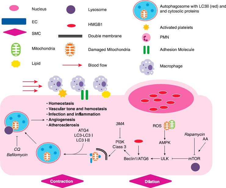
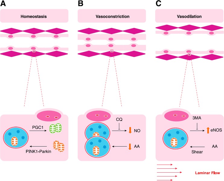
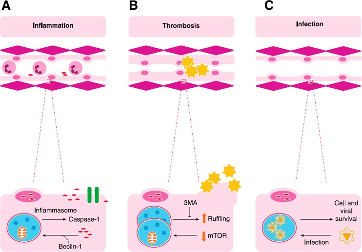
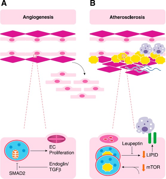
Current studies of vascular health, aging, and autophagy emphasize how the endothelium adapts to stress and contributes to disease. The endothelium is far from an inert barrier to blood-borne cells, pathogens, and chemical signals; rather, it actively translates circulating mediators into tissue responses, changing rapidly in response to physiologic stressors. Macroautophagy-the cellular ingestion of effete organelles and protein aggregates to provide anabolic substrates to fuel bioenergetics in times of stress-plays an important role in endothelial cell homeostasis, vascular remodeling, and disease. These roles include regulating vascular tone, sustaining or limiting cell survival, and contributing to the development of atherosclerosis secondary to infection, inflammation, and angiogenesis. Autophagy modulates these critical functions of the endothelium in a dynamic and perpetual response to tissue and intravascular cues.

Keywords: ATG; HMGB1; atherosclerosis; cardiovascular.


Figures

Figure 1
Figure 1
Macroautophagy in ECs. Schematic that describes pathways that mediate the induction of autophagy in ECs. Exogenous mediators known to either enhance or inhibit designated pathways are shown in italics. The common legend is utilized for all subsequent figures. Arrows designate an activating relationship. “T” lines indicate an inhibitory effect. AA, amino acids, PMN, polymorphonuclear cell, SMC, smooth muscle cell, ULK, Unc‐51–like kinase 1.
Figure 2
Figure 2
Autophagy and vascular tone. (A) Autophagic clearance of damaged mitochondria (mitophagy) is critical to maintaining homeostasis. Mitophagy is induced by PINK1‐Parkin signaling as described in the section on vascular tone, homeostasis, and the response to metabolic stress, and is balanced by mitochondrial biogenesis, induced by peroxisome proliferator‐activated receptor γ co‐activator (PGC) 1α. Effects of autophagy on ECs include modulation of NO signaling, where it has both positive and negative effects. (B and C) Whereas potassium channel ion flow is also critically important to vascular tone, these images depict autophagic regulation of NO induced vasoconstriction (B) and vasodilation (C). AA, amino acids.
Figure 3
Figure 3
EC autophagy and cell‐cell interaction. A critical role of the endothelium is modulation of cell‐cell interactions, particularly those involving leukocytes and platelets. (A) Translocation of HMGB1 from nuclei displaces beclin‐1 from BCL2, which induces autophagy. Autophagic pathways can interact with the inflammasome to release of cytokines, such as IL‐1b and IL‐18, through activation of caspase‐1. HMGB1, a nuclear protein and damage‐associated molecular pattern (DAMP) molecule, can also be released by caspase‐1–mediated pathways in Mϕs and can recruit inflammatory cells to areas of damage. (B) Typically, quiescent endothelium should not attract platelets; however, EC autophagy has been implicated in platelet recruitment as a result of cytoskeletal changes that lead to membrane ruffling. These mechanisms are thought to lead to thrombosis induced by rapamycin and sirolimus‐coated coronary stents. (C) Of interest, organisms, such as EBV, can usurp autophagic pathways to maintain a viral reservoir by promoting cell survival. Alternatively, GBS infection of cells induces autophagy, which clears bacteria, perhaps via TLR2‐mediated pathways (not shown).
Figure 4
Figure 4
Autophagy, neovascularization, and atherosclerosis. (A) EC autophagy has been implicated in angiogenesis through many mechanisms. TGF‐β and coreceptor endoglin can promote EC proliferation, a critical component of angiogenesis in which new vessels are formed from old ones, by inhibiting the inhibition of SMAD2 on autophagy. Loss of endoglin reduces autophagic markers and capillary sprouting. (B) Development of atheroma require both lipid accumulation and inflammation. Autophagosomes can eliminate lipid droplets and is thought to be promoted by the active ingredient in green tea. Leupeptin inhibits lysosomal function and prevents degradation of lipid. The accumulation of lipids within cells can promote adhesion molecule expression and Mϕ recruitment, both of which are required for atheroma formation.

References

    1. Larroque‐Cardoso, P. , Swiader, A. , Ingueneau, C. , Nègre‐Salvayre, A. , Elbaz, M. , Reyland, M. E. , Salvayre, R. , Vindis, C. (2013) Role of protein kinase C δ in ER stress and apoptosis induced by oxidized LDL in human vascular smooth muscle cells. Cell Death Dis. 4, e520. - PMC - PubMed
    1. Li, W. , Sultana, N. , Siraj, N. , Ward, L. J. , Pawlik, M. , Levy, E. , Jovinge, S. , Bengtsson, E. , Yuan, X.‐M. (2016) Autophagy dysfunction and regulatory cystatin C in macrophage death of atherosclerosis. J. Cell. Mol. Med. 20, 1664–1672. - PMC - PubMed
    1. Yuan, X.‐M. , Sultana, N. , Siraj, N. , Ward, L. J. , Ghafouri, B. , Li, W. (2016) Autophagy induction protects against 7‐oxysterol‐induced cell death via lysosomal pathway and oxidative stress. J. Cell Death 9, 1–7. - PMC - PubMed
    1. Kassiotis, C. , Ballal, K. , Wellnitz, K. , Vela, D. , Gong, M. , Salazar, R. , Frazier, O. H. , Taegtmeyer, H. (2009) Markers of autophagy are downregulated in failing human heart after mechanical unloading. Circulation 120 (Suppl 11), S191–S197. - PMC - PubMed
    1. Shirakabe, A. , Zhai, P. , Ikeda, Y. , Saito, T. , Maejima, Y. , Hsu, C.‐P. , Nomura, M. , Egashira, K. , Levine, B. , Sadoshima, J. (2016) Drp1‐dependent mitochondrial autophagy plays a protective role against pressure overload‐induced mitochondrial dysfunction and heart failure. Circulation 133, 1249–1263. - PMC - PubMed

Publication types