. 2018 Jan;32(1):176-183.
doi: 10.1038/leu.2017.193. Epub 2017 Jun 19.

Therapeutic effects of CSF1R-blocking antibodies in multiple myeloma

Affiliations

Therapeutic effects of CSF1R-blocking antibodies in multiple myeloma

Q Wang et al. Leukemia. 2018 Jan.

Abstract

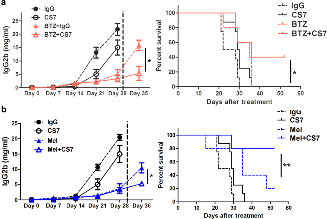
Our previous studies showed that macrophages (MФs), especially myeloma-associated MФs (MAMs), induce chemoresistance in human myeloma. Here we explored the potential of targeting MФs, by using colony-stimulating factor 1 receptor (CSF1R)-blocking mAbs, to treat myeloma. Our results showed that CSF1R blockade specifically inhibited the differentiation, proliferation and survival of murine M2 MФs and MAMs, and repolarized MAMs towards M1-like MФs in vitro. CSF1R blockade alone inhibited myeloma growth in vivo, by partially depleting MAMs, polarizing MAMs to the M1 phenotype, and inducing a tumor-specific cytotoxic CD4+ T-cell response. Similarly, genetically depleting MФs in myeloma-bearing MMDTR mice retarded myeloma growth in vivo. Furthermore, the combination of CSF1R blockade and chemotherapy such as bortezomib or melphalan displayed an additive therapeutic efficacy against established myeloma. Finally, a fully human CSF1R blocking mAb, similar to its murine counterpart, was able to inhibit the differentiation, proliferation and survival of human MФs. Thus, this study provides the first direct in vivo evidence that MΦs and MAMs are indeed important for myeloma development and progression. Our results also suggest that targeting MAMs by CSF1R blocking mAbs may be promising methods to (re)sensitize myeloma cells to chemotherapy and promote anti-myeloma immune responses in patients.


Conflict of interest statement

Conflict of Interest: The authors declare no competing financial interests.

Figures

Figure 1
Figure 1. Blocking CSF1R inhibits differentiation, survival, and proliferation of murine M2 and MAMs in vitro
(a) Viability by MTS assay in 3-day culture of CD14+ monocytes in the presence of M-CSF (CSF1) and CS7 mAb or control IgG. (b) Viability detected by MTS of murine MΦs in 3-day coculture with IL-10, IL-4, GM-CSF, LPS plus IFN-γ, or MM cells (5TGM1) in the presence of CS7 or control IgG. (c) Western blot showing the expression of phosphorylated (p) Akt and ERK in murine BMDMs cultured with M-CSF, IL-4, 5TGM1, GM-CSF, or LPS plus IFN-γ in the presence of 5 nM IgG or CS7 for 24 hours, shown is representative of at least three independent experiments. Summarized data from at least three independent experiments are shown. NS: not significant. * P<0.05; ** P<0.01; *** P<0.001.
Figure 2
Figure 2. Blocking CSF1R redirects the polarization of murine MAMs in vitro
BMDMs were cocultured with 5TGM1 in the presence of 0.5 nM CS7 (CS7) or control IgG (MAM) for 3 days. RNA (biological samples from three mice) was extracted for gene microarray and the array data were analyzed. (a) Heat maps illustrating the log2-fold change of M1 MΦ-related gene sets as indicated. (b) IPA identifying activation of specific canonical pathways as indicated, in MAMs treated with CS7 or IgG. The circle surface area is proportional to -log (p-value) and the color intensity of circles indicates the Z score. (c) Ratio in mRNA expression of indicated M1 or M2 signature genes in murine MΦs in 3-day coculture with or without 5TGM1 cells in the presence of CS4 or IgG by RT-PCR. Summarized data from at least three independent experiments are shown. NS: not significant. * P<0.05; ** P<0.01; *** P<0.001.
Figure 3
Figure 3. Blocking CSF1R inhibits murine MM growth in vivo
On day −16, KAL mice were injected with 2×10⁶ 5TGM1-luc cells via the tail vein. Starting on day 0, mice were injected intraperitoneally with 10 mg/kg CS7 (n=6 per group) or control IgG (n=6 per group) twice a week. Mouse serum was collected once a week. On day 21, mice were sacrificed for analysis. (a) Bioluminescent imaging of 5TGM1 tumor burden on day 21 (n=6 mice per group). (b) Tumor burden measured as the serum concentration of IgG2b secreted by MM cells in mice (n=6) treated with CS7 or control IgG. (c) Level of serum IgG2b on day 21 in 5TGM1-bearing MMDTR mice (n=4 per group) treated with PBS or diphtheria toxin (DT). Data are presented as mean ± SD, * P<0.05.
Figure 4
Figure 4. Blocking CSF1R induces an anti-tumor microenvironment in MM-bearing mice
Shown are numbers of MΦs in different tissues of 5TGM1-bearing KAL mice 21 days after treatment with CS7 or control IgG. (a) Dot plots showing the percentage and total number of CD11b+F4/80+ MΦs in peritoneal leukocytes, results from 1 representative mouse of total 6 mice are shown. (b) Summarized data of dot plots showing the percentage and total number of CD11b+Ly6CF4/80+ MΦs in mouse BM and spleen. (c) Fold change in the expression of M1 or M2 cytokine genes in MΦs isolated from BM of 5TGM1-bearing KAL mice (n=4) treated with CS7 or control IgG (right). Data are presented as mean ± SD. * P<0.05; ** P<0.01.
Figure 5
Figure 5. Blocking CSF1R stimulates a tumor-specific CD4+ T cell response in vivo
(a) Tumor burden of Rag−/− mice (n=9 per group) injected with 2×10⁶ 5TGM1 cells and treated with CS7 or control IgG. Tumor burden was assessed as the serum IgG2b cincentration. (b) Tumor burden detected as the circulating IgG2b concentration from 5TGM1-bearing KAL mice (n=5) treated with control IgG, CS7, CS7 plus CD8-neutralizing antibody (CS7+αCD8), CS7 plus CD4-neutralizing antibody (CS7+αCD4), or CS7 plus CD4 and CD8 neutralizing antibodies (CS7+αCD8+αCD4). (c) Representative ELISPOT images showing the number of granzyme B-secreting MM-specific CD4+ or CD8+ T cells from spleens and draining LNs of 5TGM1-bearing KAL mice treated with control IgG, CS7, CS7+αCD4, or CS7+αCD8. T cells from spleens and LNs were cocultured with mature murine DCs pulsed with or without irradiated 5TGM1 cells for 2 days before assay. Shown are results from 1 representative experiment of 4 performed (n=4 mice). Data are presented as mean ± SD of at least three independent experiments. NS: not significant.
Figure 6
Figure 6. CS7-treated MAMs promote a cytotoxic CD4+ T cell response by enhancing antigen presentation
(a) BMDMs were cocultured with irradiated 5TGM1 or 5TGM1-OVA cells in the presence of IgG or CS7 antibody for 24hs, washed four times with PBS to remove the MM cells, and then cocultured with CFSE-labeled OT-II CD4+ T cells in vitro for 2 days. Summarized results showing the percentage of CFSElow proliferative cells determined by FACS. (b) RT-PCR determination of relative Gzmb and Eomes mRNA expression in MAM-cocultured OT-II CD4+ T cells. (c) Summarized data showing the expression of CD40, CD80, CD86, and MHC II by MAMs treated with 1nM IgG or CS7 for 3 days. Data are presented as mean ± SD of at least three independent experiments. NS: not significant. * P<0.05; ** P<0.01; *** P<0.001.
Figure 7
Figure 7. The combination of CSF1R blocking mAbs and chemotherapy induces potent therapeutic efficacy of MM in vivo
(a) Tumor burden (left) and survival curves (right) of 5TGM1-bearing KAL mice (n=7 per group) treated with CS7, bortezomib (BTZ), or a combination of both. (b) Tumor burden (left) and survival curves (right) of 5TGM1-bearing KAL mice (n=7 per group) treated with CS7, melphalan (Mel), or a combination of both. Isotype IgG was used as a control for CS7. Serum samples were collected once a week. Mice were sacrificed when moribund. Tumor burden was assessed as the level of serum IgG2b. * P<0.05; ** P<0.01.
Figure 8
Figure 8. Blocking CSF1R inhibits differentiation, survival, and proliferation of human MΦs and MAM in vitro
(a) Viability in 3-day culture of human monocytes in the presence of human M-CSF (CSF1) and CS4 mAb or control IgG. (b) Viability in 3-day coculture of human MΦs with MM cells (ARP-1) in the presence of CS4 or control IgG. (c) Summarized data showing the percentage of Ki67+CD11b+ proliferating MΦs in 24-hour coculture with MM cells (ARP-1) in the presence of CS4 or control IgG. Summarized data from three independent experiments are shown. NS: not significant. * P<0.05; ** P<0.01.

References

    1. Hideshima T, Mitsiades C, Tonon G, Richardson PG, Anderson KC. Understanding multiple myeloma pathogenesis in the bone marrow to identify new therapeutic targets. Nat Rev Cancer. 2007 Aug;7(8):585–598. - PubMed
    1. Podar K, Chauhan D, Anderson KC. Bone marrow microenvironment and the identification of new targets for myeloma therapy. Leukemia. 2009 Jan;23(1):10–24. - PMC - PubMed
    1. Gordon S, Taylor PR. Monocyte and macrophage heterogeneity. Nature reviews Immunology. 2005 Dec;5(12):953–964. - PubMed
    1. Wynn TA, Chawla A, Pollard JW. Macrophage biology in development, homeostasis and disease. Nature. 2013 Apr 25;496(7446):445–455. - PMC - PubMed
    1. Biswas SK, Mantovani A. Macrophage plasticity and interaction with lymphocyte subsets: cancer as a paradigm. Nature immunology. 2010 Oct;11(10):889–896. - PubMed

Publication types

MeSH terms