. 2017:2017:8398072.
doi: 10.1155/2017/8398072. Epub 2017 May 24.

The Effects of Blast Exposure on Protein Deimination in the Brain

Affiliations

The Effects of Blast Exposure on Protein Deimination in the Brain

Peter J Attilio et al. Oxid Med Cell Longev. 2017.

Abstract

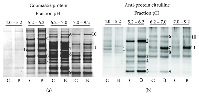
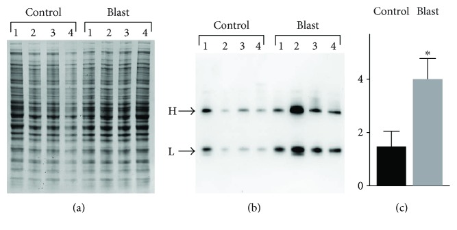
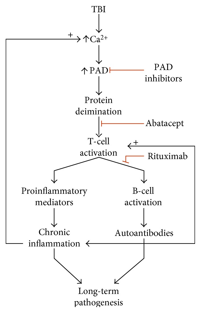
Oxidative stress and calcium excitotoxicity are hallmarks of traumatic brain injury (TBI). While these early disruptions may be corrected over a relatively short period of time, long-lasting consequences of TBI including impaired cognition and mood imbalances can persist for years, even in the absence of any evidence of overt injury based on neuroimaging. This investigation examined the possibility that disordered protein deimination occurs as a result of TBI and may thus contribute to the long-term pathologies of TBI. Protein deimination is a calcium-activated, posttranslational modification implicated in the autoimmune diseases rheumatoid arthritis and multiple sclerosis, where aberrant deimination creates antigenic epitopes that elicit an autoimmune attack. The present study utilized proteomic analyses to show that blast TBI alters the deimination status of proteins in the porcine cerebral cortex. The affected proteins represent a small subset of the entire brain proteome and include glial fibrillary acidic protein and vimentin, proteins reported to be involved in autoimmune-based pathologies. The data also indicate that blast injury is associated with an increase in immunoglobulins in the brain, possibly representing autoantibodies directed against novel protein epitopes. These findings indicate that aberrant protein deimination is a biomarker for blast TBI and may therefore underlie chronic neuropathologies of head injury.


Figures

Figure 1
Figure 1
Protein deimination is catalyzed by a family of structurally related, calcium-dependent enzymes known as peptidylarginine deiminases (PADs). Protein deimination involves the conversion of an intraprotein arginine residue to a citrulline residue, resulting in the loss of a positively charged amine group and 1 Da in molecular mass.
Figure 2
Figure 2
Blast-induced deimination of proteins in porcine brain. Brain samples were collected 2 weeks following a single blast exposure (average pressure = 46 psi). Homogenates of control (C) and the blast-exposed (B) cerebral cortex were prefractionated by LP-IEF. The resulting pH fractions were further fractionated by 1-dimensional SDS-PAGE (a) and analyzed for protein deimination by western blotting (b) using the mouse monoclonal 6B3 anti-protein citrulline antibody. Immunoreactive features affected by TBI (numbered, panel (b)) were mapped to corresponding bands in a Coomassie-stained protein gel (numbered, panel (a)). These were collected, identified, and mapped for site-specific deimination by peptide mass fingerprinting using liquid chromatography and tandem mass spectrometry (LC MS/MS).
Figure 3
Figure 3
Mapping of protein deimination sites by neutral loss. Tryptic peptides were fragmented by collision-induced dissociation, and resulting spectra were analyzed for a neutral loss of 43 Da, reflecting the loss of isocyanic acid as a fragmentation product of citrulline (upper panel). The representative spectrum shown here depicts the Y and B ion spectra of GFAP peptide, TVEMrDGEVIK, with the neutral loss peak observed for the deiminated arginine (r) at 625.8 Da. Because the parent peptide ion was doubly charged in this case, the observed neutral loss in the spectrum was 21.5 Da, reflecting 43 Da/2.
Figure 4
Figure 4
Effects of blast exposure on the presence of IgG expression in the cerebral cortex of swine. Homogenates of control and the blast-exposed (2 weeks postinjury) cerebral cortex (N-4/group) were fractionated by (a) 1-dimensional SDS-PAGE and (b) analyzed for IgG content by western blotting. Immunoreactive heavy (H) and light (L) chain IgGs were visualized using an anti-porcine IgG detection antibody. The values for the total IgG chemiluminescence signal (H+L) (c) for each sample were standardized to protein load (a) by densitometry analysis using ImageJ, and resulting values were analyzed statistically. The relative signal intensity is shown on the y-axis as densitometry values ×100. Data are presented as the mean ± standard error; p ≤ 0.005.
Figure 5
Figure 5
Proposed mechanism for the role of aberrant protein deimination in an autoimmune response to brain injury. TBI-induced calcium excitotoxicity hyperactivates PAD resulting in an abnormal pattern of protein deimination. Cells of the adaptive immune system process the modified proteins to reveal antigenic epitopes created by deimination. Antigen presentation and T-cell activation subsequently lead to the activation of B-cells for the production of autoantibodies and chronic neuroinflammation. It is proposed that these mechanisms contribute to long-term pathologies that can result from TBI. Potential therapeutic interventions that inhibit protein deimination and T-cell and B-cell activation are depicted with red lines.

References

    1. Ansari M. A., Roberts K. N., Scheff S. W. Oxidative stress and modification of synaptic proteins in hippocampus after traumatic brain injury. Free Radical Biology & Medicine. 2008;45(2):443–452. doi: 10.1016/j.freeradbiomed.2008.04.038. - DOI - PMC - PubMed
    1. Darwish R. S., Amiridze N., Aarabi B. Nitrotyrosine as an oxidative stress marker: evidence for involvement in neurologic outcome in human traumatic brain injury. The Journal of Trauma. 2007;63(2):439–442. doi: 10.1097/TA.0b013e318069178a. - DOI - PubMed
    1. Goldstein L. E., Fisher A. M., Tagge C. A., et al. Chronic traumatic encephalopathy in blast-exposed military veterans and a blast neurotrauma mouse model. Science Translational Medicine. 2012;4(134):p. 134ra160. doi: 10.7812/TPP/16-055. - DOI - PMC - PubMed
    1. Opii W. O., Nukala V. N., Sultana R., et al. Proteomic identification of oxidized mitochondrial proteins following experimental traumatic brain injury. Journal of Neurotrauma. 2007;24(5):772–789. doi: 10.1089/neu.2006.0229. - DOI - PubMed
    1. de Lanerolle N. C., Bandak F., Kang D., et al. Characteristics of an explosive blast-induced brain injury in an experimental model. Journal of Neuropathology and Experimental Neurology. 2011;70(11):1046–1057. doi: 10.1097/NEN.0b013e318235bef2. - DOI - PubMed