Review
. 2017 Oct;1862(10 Pt B):1166-1177.
doi: 10.1016/j.bbalip.2017.06.006. Epub 2017 Jun 13.

Establishing the lipid droplet proteome: Mechanisms of lipid droplet protein targeting and degradation

Affiliations
Review

Establishing the lipid droplet proteome: Mechanisms of lipid droplet protein targeting and degradation

Kirill Bersuker et al. Biochim Biophys Acta Mol Cell Biol Lipids. 2017 Oct.

Abstract

Lipid droplets (LDs) are ubiquitous, endoplasmic reticulum (ER)-derived organelles that mediate the sequestration of neutral lipids (e.g. triacylglycerol and sterol esters), providing a dynamic cellular storage depot for rapid lipid mobilization in response to increased cellular demands. LDs have a unique ultrastructure, consisting of a core of neutral lipids encircled by a phospholipid monolayer that is decorated with integral and peripheral proteins. The LD proteome contains numerous lipid metabolic enzymes, regulatory scaffold proteins, proteins involved in LD clustering and fusion, and other proteins of unknown functions. The cellular role of LDs is inherently determined by the composition of its proteome and alteration of the LD protein coat provides a powerful mechanism to adapt LDs to fluctuating metabolic states. Here, we review the current understanding of the molecular mechanisms that govern LD protein targeting and degradation. This article is part of a Special Issue entitled: Recent Advances in Lipid Droplet Biology edited by Rosalind Coleman and Matthijs Hesselink.

Keywords: Lipid droplet; Membrane protein; Protein degradation; Protein targeting; Proteome; Ubiquitin.


Figures

Fig. 1
Fig. 1
The LD proteome: Structural features and targeting pathways. Proteins utilize a variety of mechanisms for association with LDs, including insertion into the membrane via hydrophobic hairpin structures (Class I), amphipathic helices (Class II), association with the membrane via lipid modifications, and indirect recruitment via interactions with integral proteins. Class I proteins insert into the ER and are subsequently trafficked to LDs.
Fig. 2
Fig. 2
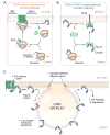
Mechanisms of LD protein insertion into the ER and ER-LD protein sorting. (A) Two pathways have been implicated in the insertion of Class I LD proteins into the ER. The canonical SRP-Sec61 pathway (red box) mediates the insertion of caveolin-1 and oleosin. In this pathway, SRP recognizes the hydrophobic region as it emerges from the ribosome and translation is stalled. SRP docks with the ER-resident SRP receptor, facilitating ribosome association with the Sec61 translocon, and the proteins are cotranslationally inserted into the ER. (B) The PEX19-PEX3 pathway (blue box) mediates the insertion of UBXD8. PEX19 binds the hydrophobic hairpin region of UBXD8. Through association with its receptor PEX3, farnesylated PEX19 then mediates the posttranslational insertion of UBXD8 into discrete ER subdomains. (C) Multiple mechanisms contribute to the sorting of ER and LD proteomes, including 1) a topology-selective diffusion barrier formed by the junction of the LD monolayer and the ER bilayer membranes, 2) protein interactions with ER-resident proteins, which function as tethers and impact ER motility, 3) protein domains that have an inherent preference for the LD monolayer, and 4) instability in the ER, facilitating enrichment in LDs.
Fig. 3
Fig. 3
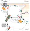
Potential routes for LD protein degradation by the ubiquitin-proteasome system. Emerging evidence suggests that LD proteins (shown in green) can be degraded by multiple ubiquitin-proteasome pathways under different conditions. In the absence of LDs, LD proteins are likely degraded by both ER ubiquitination pathways (i.e. ERAD) and cytosolic ubiquitination pathways. The presence of LD-localized ubiquitination components suggests the existence of a LD-associated ubiquitination pathway.

References

    1. Walther TC, Farese RV. Lipid droplets and cellular lipid metabolism. Annu Rev Biochem. 2012;81:687–714. doi: 10.1146/annurev-biochem-061009-102430. - DOI - PMC - PubMed
    1. Pol A, Gross SP, Parton RG. Review: biogenesis of the multifunctional lipid droplet: lipids, proteins, and sites. J Cell Biol. 2014;204:635–646. doi: 10.1083/jcb.201311051. - DOI - PMC - PubMed
    1. Hashemi HF, Goodman JM. The life cycle of lipid droplets. Curr Opin Cell Biol. 2015;33:119–124. doi: 10.1016/j.ceb.2015.02.002. - DOI - PMC - PubMed
    1. Gao Q, Goodman JM. The lipid droplet-a well-connected organelle. Front Cell Dev Biol. 2015;3:49. doi: 10.3389/fcell.2015.00049. - DOI - PMC - PubMed
    1. Listenberger LL, Han X, Lewis SE, Cases S, Farese RV, Ory DS, et al. Triglyceride accumulation protects against fatty acid-induced lipotoxicity. Proc Natl Acad Sci U S A. 2003;100:3077–3082. doi: 10.1073/pnas.0630588100. - DOI - PMC - PubMed

Publication types