Review
. 2017 Aug;38(2):665-675.
doi: 10.3892/or.2017.5714. Epub 2017 Jun 13.

Exosomes: New players in cancer (Review)

Affiliations
Review

Exosomes: New players in cancer (Review)

Wei Guo et al. Oncol Rep. 2017 Aug.

Abstract

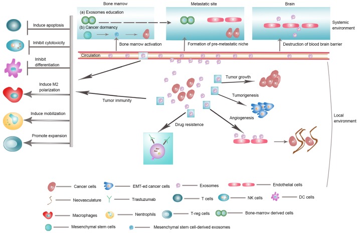
The past decade has witnessed an exponential increase in research on exosomes. For many years considered to be extracellular debris, exosomes are now considered important mediators in intercellular communication. The capability of exosomes to transfer proteins, DNA, mRNA, as well as non-coding RNAs has made them an attractive focus of research into the pathogenesis of different diseases, including cancer. Increasing evidence suggests that tumor cells release a large sum of exosomes, which may not only influence proximal tumor cells and stromal cells in local microenvironment, but also can exert systemic effects when participating in blood circulation. In this study, we review the current understanding on this topic. The literature outlines two broad facets of exosomes in cancer: 1) promotion of tumor growth, tumorigenesis, tumor angiogenesis, tumor immune escape, drug resistance, and metastasis and 2) their role as promising biomarkers for cancer diagnosis and even as potential treatment targets for cancer patients.


Figures

Figure 1.
Figure 1.
Biogenesis, release, content and uptake of exosomes. Early endosome is formed from the plasma membrane via endocytic pathway. MVB can be formed by the invagination of endosomal membrane. Dependent on the function and content, MVB then can be directed to fuse with plasma membrane and release to the extracellular space as exosomes. During the biogenesis of exosomes and prior to their secretion, proteins (e.g., tetraspanin, cytosolic proteins, receptor), nucleic acids (e.g., mRNA, miRNA, DNA), and lipids (e.g., sphingomyelin, cholesterol) are uploaded to exosomes. Cells appear to take up exosomes via several ways: (a) receptor-/lipidraft medated endocytosis, (b) phagocytosis, (c) macropinocytosis, (d) fusion with the plasma membrane of the target cell. MVB: multivesicular body.
Figure 2.
Figure 2.
Biological function of exosomes in cancer. Exosomes are involved in tumor growth, tumorigenesis, angiogenesis, tumor immune escape, drug resistance and metastasis. EMT, epithelial-mesenchymal transition.

References

    1. Trams EG, Lauter CJ, Salem N, Jr, Heine U. Exfoliation of membrane ecto-enzymes in the form of micro-vesicles. Biochim Biophys Acta. 1981;645:63–70. doi: 10.1016/0005-2736(81)90512-5. - DOI - PubMed
    1. Harding C, Heuser J, Stahl P. Receptor-mediated endocytosis of transferrin and recycling of the transferrin receptor in rat reticulocytes. J Cell Biol. 1983;97:329–339. doi: 10.1083/jcb.97.2.329. - DOI - PMC - PubMed
    1. Fong MY, Zhou W, Liu L, Alontaga AY, Chandra M, Ashby J, Chow A, O'Connor ST, Li S, Chin AR, et al. Breast-cancer-secreted miR-122 reprograms glucose metabolism in premetastatic niche to promote metastasis. Nat Cell Biol. 2015;17:183–194. doi: 10.1038/ncb3094. - DOI - PMC - PubMed
    1. Peinado H, Aleckovic M, Lavotshkin S, Matei I, Costa-Silva B, Moreno-Bueno G, Hergueta-Redondo M, Williams C, García-Santos G, Ghajar CM, et al. Melanoma exosomes educate bone marrow progenitor cells toward a pro-metastatic phenotype through MET. Nat Med. 2012;18:883–891. doi: 10.1038/nm.2753. - DOI - PMC - PubMed
    1. Li J, Liu K, Liu Y, Xu Y, Zhang F, Yang H, Liu J, Pan T, Chen J, Wu M, et al. Exosomes mediate the cell-to-cell transmission of IFN-[alpha]-induced antiviral activity. Nat Immunol. 2013;14:793–803. doi: 10.1038/ni.2647. - DOI - PubMed

Substances