. 2017 Jul 11;56(27):3507-3517.
doi: 10.1021/acs.biochem.7b00268. Epub 2017 Jun 30.

The Sulfur-Linked Analogue of O-GlcNAc (S-GlcNAc) Is an Enzymatically Stable and Reasonable Structural Surrogate for O-GlcNAc at the Peptide and Protein Levels

Affiliations

The Sulfur-Linked Analogue of O-GlcNAc (S-GlcNAc) Is an Enzymatically Stable and Reasonable Structural Surrogate for O-GlcNAc at the Peptide and Protein Levels

Cesar A De Leon et al. Biochemistry. .

Abstract

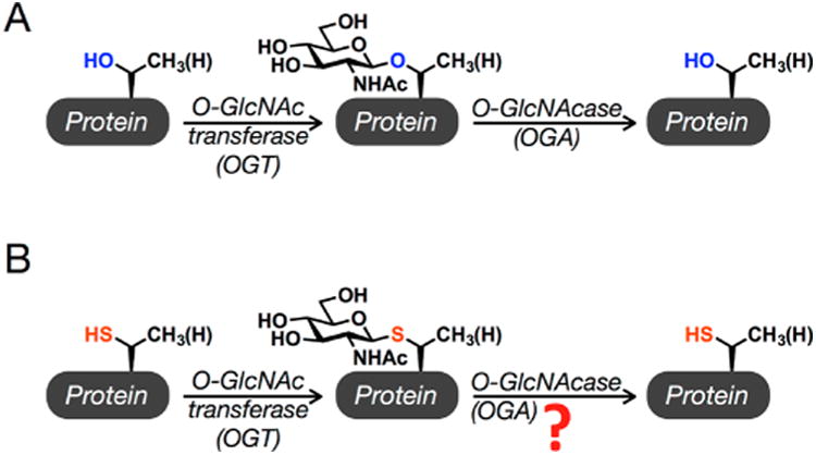
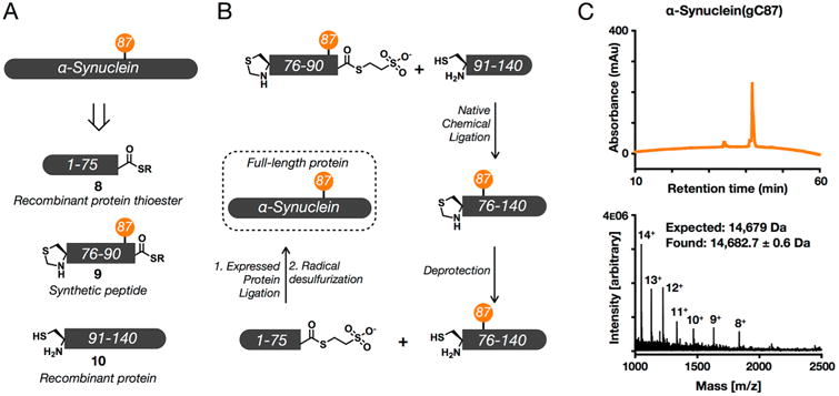
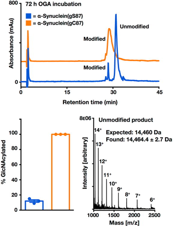
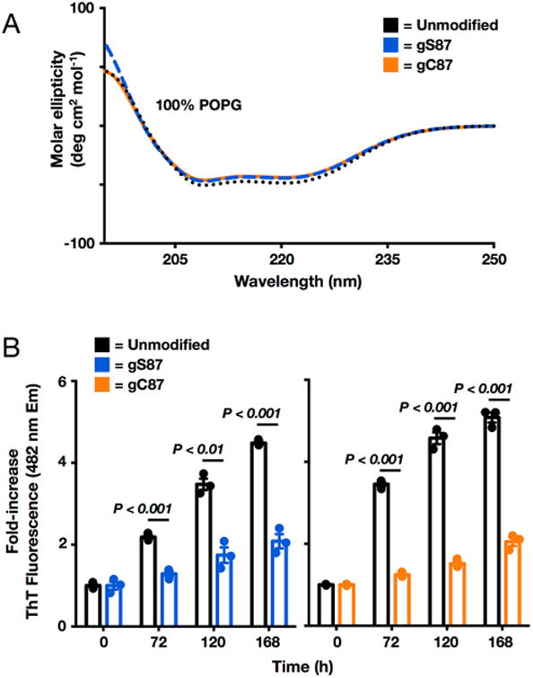
Synthetic proteins bearing site-specific posttranslational modifications have revolutionized our understanding of their biological functions in vitro and in vivo. One such modification, O-GlcNAcylation, is the dynamic addition of β-N-acetyl glucosamine to the side chains of serine and threonine residues of proteins, yet our understanding of the site-specific impact of O-GlcNAcylation remains difficult to evaluate in vivo because of the potential for enzymatic removal by endogenous O-GlcNAcase (OGA). Thioglycosides are generally perceived to be enzymatically stable structural mimics of O-GlcNAc; however, in vitro experiments with small-molecule GlcNAc thioglycosides have demonstrated that OGA can hydrolyze these linkages, indicating that S-linked β-N-acetyl glucosamine (S-GlcNAc) on peptides or proteins may not be completely stable. Here, we first develop a robust synthetic route to access an S-GlcNAcylated cysteine building block for peptide and protein synthesis. Using this modified amino acid, we establish that S-GlcNAc is an enzymatically stable surrogate for O-GlcNAcylation in its native protein setting. We also applied nuclear magnetic resonance spectroscopy and computational modeling to find that S-GlcNAc is an good structural mimic of O-GlcNAc. Finally, we demonstrate that site-specific S-GlcNAcylation results in biophysical characteristics that are the same as those of O-GlcNAc within the context of the protein α-synuclein. While this study is limited in focus to two model systems, these data suggest that S-GlcNAc broadly resembles O-GlcNAc and that it is indeed a stable analogue in the context of peptides and proteins.


Figures

Figure 1
Figure 1
O-GlcNAcylation and the corresponding S-GlcNAc analogue. (A) O-GlcNAcylation is the addition of N-acetyl glucos-amine to serine and threonine residues of intracellular proteins. It is added by the enzyme O-GlcNAc transferase (OGT) and removed by O-GlcNAcase (OGA). (B) S-GlcNAc analogues of O-GlcNAc could be used as enzymatically stable analogues in synthetic proteins, but whether human OGA can remove these modifications was an open question.
Figure 2
Figure 2
Synthetic routes to S-GlcNAcylated amino acids for solid-phase peptide synthesis. (A) Previous routes involved nucleophilic displacement of leaving groups (LG) on alanine derivatives. (B) The new synthetic route to S-GlcNAcylated cysteine developed here. Reagents: (a) 20 mol % InBr3, Fmoc-Cys-OH, CH2Cl2, reflux, 1–16 h; (b) 20 mol % InBr3, Fmoc-Cys-OH, dichloroethane, reflux, 16 h, 90%; (c) Zn dust, AcOH, Ac2O, 16 h; (d) pentafluorophenyl trifluoroacetate, pyridine, 3 h, 91%; (e) Zn dust, AcOH, Ac2O, 16 h, 70%.
Figure 3
Figure 3
S-GlcNAc is a good structural mimic of O-GlcNAc. Comparison of ab initio folding, QM modeling, and backbone HN and Hα chemical shift deviations (CSDs) for model O/S-GlcNAc-modified peptides. Ab initio folding was conducted within the Rosetta Molecular Design package (see the Supporting Information for “folding funnels” and O/S GlcNAc residue parametrizations). The lowest-energy structure from the ab initio folding is presented. The β-hairpin conformation observed in the lowest-energy ab initio modeling was extracted and geometrically optimized at the B3LYP/6-311+G-(p,2d) level of theory.
Figure 4
Figure 4
In the context of a peptide, S-GlcNAc is completely stable against human OGA enzymatic deglycosylation. O-GlcNAcylated (blue) or S-GlcNAcylated (orange) peptides (50 μM, in PBS at pH 7.4) were incubated with human OGA (1 μM) at 37 °C for ≤72 h. The S-GlcNAcylated peptide HPLC trace is offset in the y-direction for the sake of clarity. mAU indicates milliabsorbance units. RP-HPLC conditions were 0–70% buffer B over 60 min; buffer A consisted of 0.1% TFA in H2O, and buffer B consisted of 0.1% TFA and 90% ACN in H2O.
Figure 5
Figure 5
Semisynthesis of S-GlcNAcylated α-synuclein. (A) α-Synuclein was retrosynthetically deconstructed into a recombinant protein thioester (8) obtained using intein chemistry, a synthetic thioester peptide (9), and a recombinant protein (10). (B) These fragments were then combined through iterative ligation reactions. (C) Characterization of synthetic S-GlcNAcylated α-synuclein using RP-HPLC and electrospray ionization mass spectrometry (ESI-MS). Analysis by RP-HPLC showed that synthetic α-synuclein(gC87) was pure, as evidenced by the appearance of only one sharp peak. Characterization by ESI-MS gave a range of charge states that could be deconvoluted to a molecular mass (14682.7 ± 0.6 Da) in excellent agreement with the predicted weight of 14679 Da. RP-HPLC conditions were 0–70% buffer B over 60 min; buffer A consisted of 0.1% TFA in H2O, and buffer B consisted of 0.1% TFA and 90% ACN in H2O.
Figure 6
Figure 6
S-GlcNAcylation of α-synuclein is enzymatically stable. α-Synuclein(gS87) or α-synuclein(gC87) (25 μM, in PBS at pH 7.4) was incubated in triplicate with OGA (1 μM) at 37 °C for 72 h. S-GlcNAc is offset in the y-direction for the sake of clarity. mAU indicates milliabsorbance units. Hydrolysis of GlcNAcylated α-synuclein was analyzed by HPLC at the 72 h time point. Deglycosylation was quantitated by area percent using high-performance liquid chromatography (HPLC) at 214 nm, and the deglycosylated product was characterized by ESI-MS. Results are the mean ± the standard error of the mean of three separate biological experiments. RP-HPLC conditions were 35–60% buffer B over 60 min; buffer A consisted of 0.1% TFA in H2O, and buffer B consisted of 0.1% TFA and 90% ACN in H2O.
Figure 7
Figure 7
S-GlcNAcylation has effects identical to those of O-GlcNAcylation on the membrane binding and aggregation of α-synuclein. (A) Neither O-GlcNAcylation nor S-GlcNAcylation at residue 87 affects α-synuclein membrane binding. Recombinant α-synuclein, α-synuclein(gS87), or α-synuclein(gC87) was incubated with a 100-fold excess of POPG preformed vesicles and analyzed using circular dichroism (CD). All of the proteins gave essentially indistinguishable CD spectra consistent with the formation of an extended α-helix. POPG denotes 1-palmitoyl-2-oleoyl-sn-glycero-3-[phospho-rac-(1-glycerol)]. (B) O-GlcNAcylation and S-GlcNAcylation are equally inhibitory toward a-synuclein aggregation. Recombinant α-synuclein, α-synuclein(gS87), or α-synuclein(gC87) was subjected to aggregation conditions (25 μM concentration and agitation at 37 °C) for the indicated lengths of time before analysis of aliquots by ThT fluorescence (λex = 450 nm, and λem = 482 nm). The y-axis shows the fold increase in fluorescence compared with the corresponding protein at time zero. Error bars represent the standard error of the mean from the mean of three biological replicates, and statistical significance was calculated using a two-tailed Student's t test.

References

    1. Bond MR, Hanover JA. A little sugar goes a long way: the cell biology of O-GlcNAc. J Cell Biol. 2015;208:869–880. - PMC - PubMed
    1. Vocadlo DJ. O-GlcNAc processing enzymes: catalytic mechanisms, substrate specificity, and enzyme regulation. Curr Opin Chem Biol. 2012;16:488–497. - PubMed
    1. Shafi R, Iyer SP, Ellies LG, O'Donnell N, Marek KW, Chui D, Hart GW, Marth JD. The O-GlcNAc transferase gene resides on the X chromosome and is essential for embryonic stem cell viability and mouse ontogeny. Proc Natl Acad Sci U S A. 2000;97:5735–5739. - PMC - PubMed
    1. O'Donnell N, Zachara NE, Hart GW, Marth JD. Ogt-dependent X-chromosome-linked protein glycosylation is a requisite modification in somatic cell function and embryo viability. Mol Cell Biol. 2004;24:1680–1690. - PMC - PubMed
    1. Sinclair DAR, Syrzycka M, Macauley MS, Rastgardani T, Komljenovic I, Vocadlo DJ, Brock HW, Honda BM. Drosophila O-GlcNAc transferase (OGT) is encoded by the Polycomb group (PcG) gene, super sex combs (sxc) Proc Natl Acad Sci U S A. 2009;106:13427–13432. - PMC - PubMed

Publication types

MeSH terms