Case Reports
. 2017 Jun;30(3):235-239.
doi: 10.1177/1971400917695318. Epub 2017 Mar 13.

Pharyngeal amyloidomas: Variable appearance on imaging

Affiliations
Case Reports

Pharyngeal amyloidomas: Variable appearance on imaging

C A Prause et al. Neuroradiol J. 2017 Jun.

Abstract

Amyloidomas are rare tumor-like depositions of abnormally folded, insoluble proteins that may be seen in the setting of systemic amyloidosis or as isolated tumoral deposits. Focal, isolated amyloidomas carry an excellent prognosis whereas systemic amyloidoses do not. The ability to identify or suggest amyloidoma on imaging studies may help direct laboratory testing and eventual diagnosis. Amyloidomas involving the head and neck have been variably described from homogeneously T2 hypointense to iso-slightly hyperintense relative to skeletal muscle. Herein we present two patients with pharyngeal submucosal amyloidomas of differing sizes and imaging characteristics to emphasize their potential widely variable imaging appearance and broaden our knowledge of these rare lesions.

Keywords: Amyloidomas; amyloidoses; amyloidosis; head and neck; plasmacytoma.


Figures

Figure 1.
Figure 1.
A 55-year-old female with bilateral retropharyngeal amyloidomas. PET/CT coronal image (a) demonstrates heterogeneous FDG uptake, with greatest FDG uptake within the amyloidomas’ upper nasopharyngeal component (thick arrows) and geographic regions of low FDG uptake within the lower lesions (thin arrows) at the oropharynx level. Contrast-enhanced neck CT axial image (b) at the upper oropharynx level reveals well-defined bilateral retropharyngeal masses, containing tiny calcifications on the left (small black arrow). On CT these masses demonstrate variable contrast enhancement that is more homogeneous and intense on the right (thick white arrow) than the left (thin white arrow). MRI coronal T2 (c), T1 (d) and post-contrast T1 (e)-weighted images demonstrate amyloidoma heterogeneity, with their upper FDG avid component appearing T2 and mildly T1 hyperintense relative to muscle with marked post-contrast enhancement (thick arrows). The inferior amyloidoma components (thin arrows), with predominant low FDG uptake, are mildly T2 and T1 hypointense to muscle, with heterogeneous, less intense contrast enhancement. Pathology slide (H/E stain, 10× magnification) (f) from oropharynx level biopsy of the right pharyngeal lesion demonstrates a heavy background of eosinophilia (black arrow) with scant plasma cells, and a focal collection of plasma cells within its superior aspect (thick white arrow). PET/CT: positron emission tomography/computed tomography; FDG: F-18 fluorodeoxyglucose; MRI: magnetic resonance imaging; H/E: hematoxylin and eosin.
Figure 2.
Figure 2.
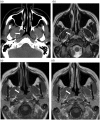
A 48-year-old female with nasopharyngeal submucosal amyloidoma. Contrast-enhanced CT axial image (a) reveals a submucosal non-enhancing lesion (thick arrow) containing two small calcifications (thin arrow) contiguous with an enlarged torus tubarius. MRI axial T2 (b), T1 (c) and post-contrast T1-weighted (d) images demonstrate the amyloidoma (arrow) relative to muscle to be T2 hyperintense, T1 isointense and non-enhancing. CT: computed tomography; MRI: magnetic resonance imaging.

References

    1. Parmar H, Rath T, Castillo M, et al. Imaging of focal amyloid depositions in the head, neck, and spine: Amyloidoma. AJNR Am J Neuroradiol 2010; 31: 1165–1170. - PMC - PubMed
    1. Penner C, Muller S. Head and neck amyloidosis: A clinicopathologic study of 15 cases. Oral Oncol 2006; 42: 421–429. - PubMed
    1. Falk R, Comenzo R, Skinner M. The systemic amyloidoses. New Engl J Med 1997; 337: 898–909. - PubMed
    1. Bardin RL, Barnes CE, Stanton CA, et al. Soft tissue amyloidoma of the extremities: A case report and review of the literature. Arch Pathol Lab Med 2004; 128: 1270–1273. - PubMed
    1. Pietruszewska W, Wagrowska-Danilewicz M, Klatka J. Amyloidosis of the head and neck: A clinicopatholgical study of cases with long-term follow-up. Arch Med Sci 2014; 10: 846–852. - PMC - PubMed

Publication types

LinkOut - more resources