. 2017 Dec;14(6):066005.
doi: 10.1088/1741-2552/aa7a42.

A novel flexible cuff-like microelectrode for dual purpose, acute and chronic electrical interfacing with the mouse cervical vagus nerve

Affiliations

A novel flexible cuff-like microelectrode for dual purpose, acute and chronic electrical interfacing with the mouse cervical vagus nerve

A S Caravaca et al. J Neural Eng. 2017 Dec.

Abstract

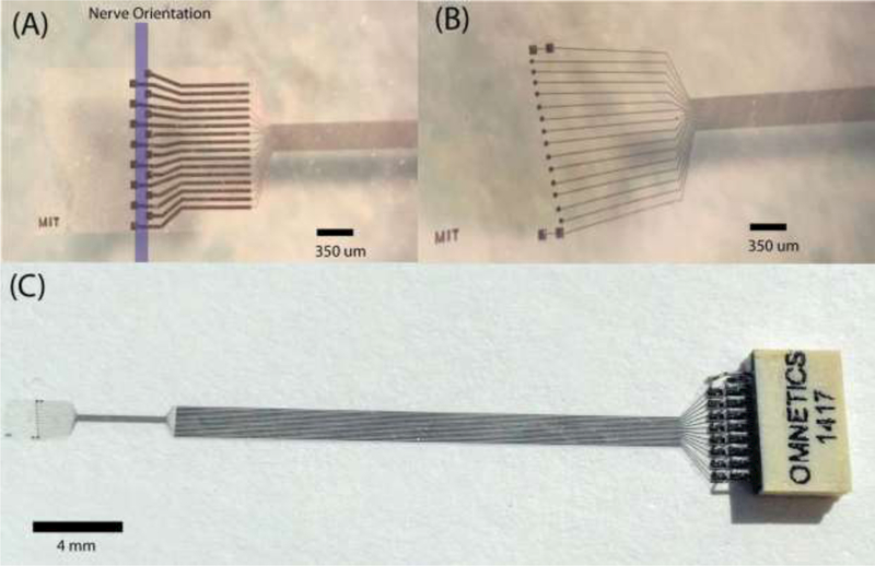
Objective: Neural reflexes regulate immune responses and homeostasis. Advances in bioelectronic medicine indicate that electrical stimulation of the vagus nerve can be used to treat inflammatory disease, yet the understanding of neural signals that regulate inflammation is incomplete. Current interfaces with the vagus nerve do not permit effective chronic stimulation or recording in mouse models, which is vital to studying the molecular and neurophysiological mechanisms that control inflammation homeostasis in health and disease. We developed an implantable, dual purpose, multi-channel, flexible 'microelectrode' array, for recording and stimulation of the mouse vagus nerve.

Approach: The array was microfabricated on an 8 µm layer of highly biocompatible parylene configured with 16 sites. The microelectrode was evaluated by studying the recording and stimulation performance. Mice were chronically implanted with devices for up to 12 weeks.

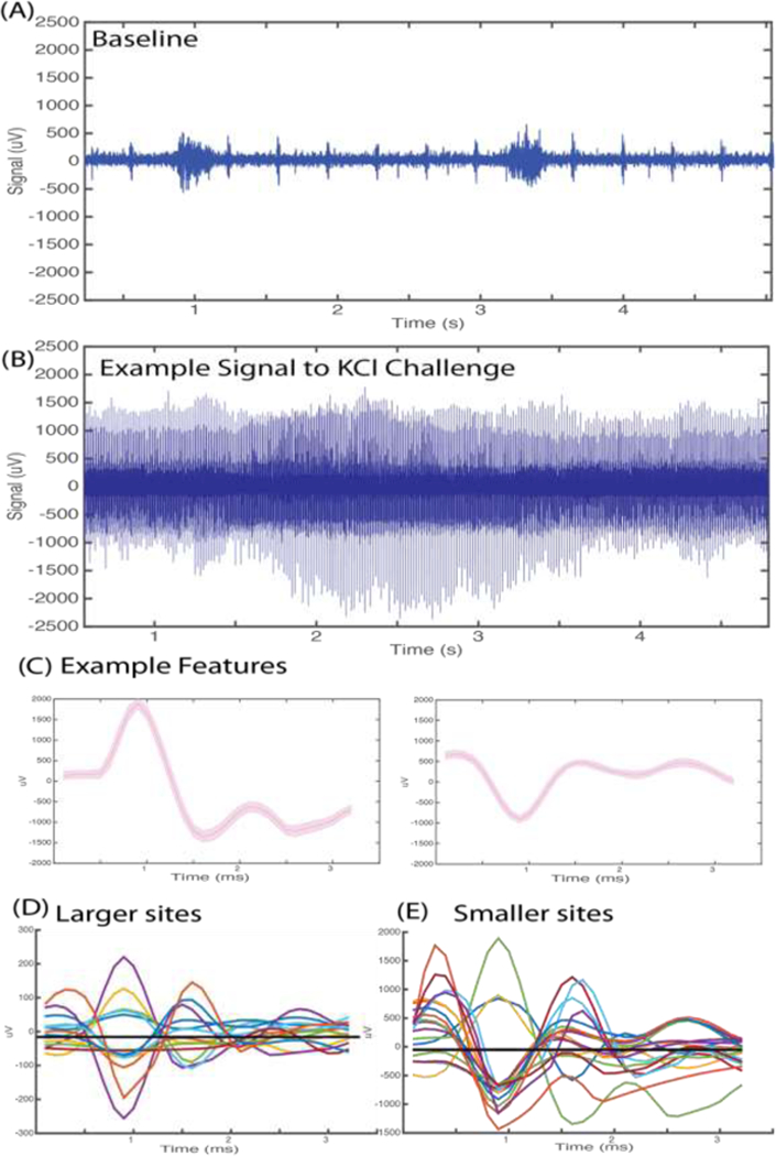
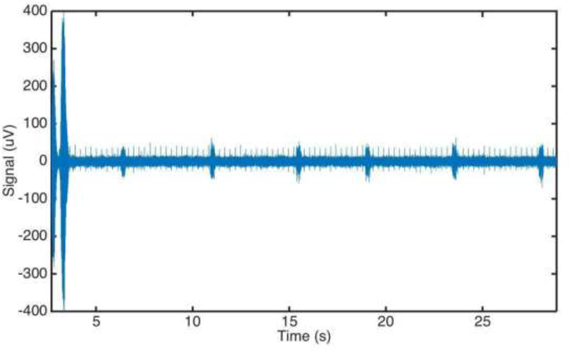
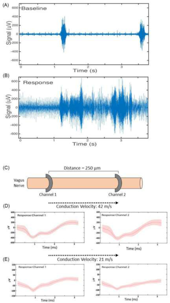
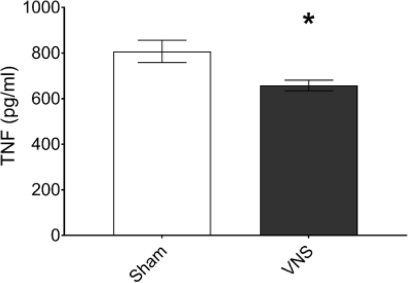
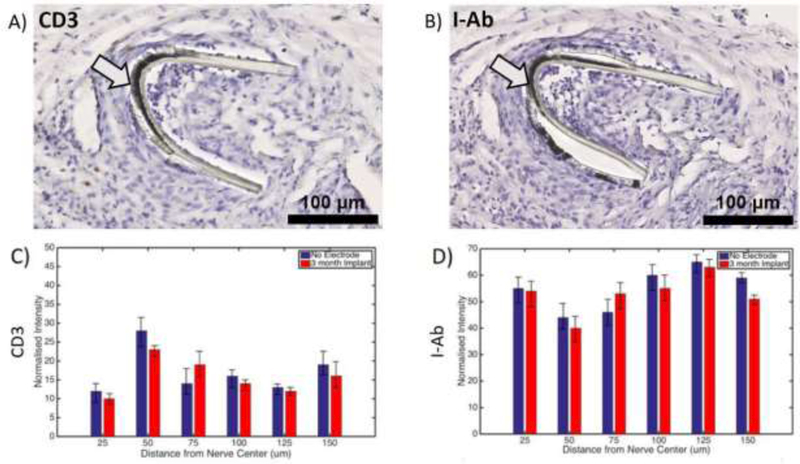
Main results: Using the microelectrode in vivo, high fidelity signals were recorded during physiological challenges (e.g potassium chloride and interleukin-1β), and electrical stimulation of the vagus nerve produced the expected significant reduction of blood levels of tumor necrosis factor (TNF) in endotoxemia. Inflammatory cell infiltration at the microelectrode 12 weeks of implantation was limited according to radial distribution analysis of inflammatory cells.

Significance: This novel device provides an important step towards a viable chronic interface for cervical vagus nerve stimulation and recording in mice.


Figures

Figure 1.
Figure 1.
Microelectrode designs for the peripheral implant in the mouse. (a) A tetrode like electrode format with the direction of the nerve, recording site area 6400 μm2. The oversized flap wraps around the nerve and is secured in place with the use of sutures or surgically approved adhesives. (b) The diagonal format of recording sites (2500 μm2) with two macro stimulation sites (6400 μm2) at either end. (c) The microelectrode shown with incorporated ribbon cable and connector.
Figure 2.
Figure 2.
Electrical vagus nerve activity in response to potassium chloride (KCI). Anesthetized mice were implanted with a microelectrode on the left cervical vagus nerve. Data was continuously acquired at 30 kHz sampling rate. (A) Example of 200 Hz high passed signal of recorded baseline activity. Burst activity synchronous with pulse and respiration was observed. (B) Example of 200 Hz high passed signal of vagus nerve activity upon cervical vagus nerve exposure to 4 mM KCI. (C) Representative features extracted from the raw signal recording upon KCI challenge. (D) Mean waveforms of CAPs extracted from activity recorded at the 6400 μm2 recording sites. (E) Mean waveforms of CAPs extracted from activity recorded at 2500 μm2 recording sites.
Figure 3.
Figure 3.
Electrical cervical vagus nerve activity following intraperitoneal injection of saline. Anesthetized mice were implanted with a microelectrode on the left cervical vagus nerve. Data was continuously acquired at 30 kHz sampling rate. Injection was performed at time ‘0’. A movement artifact from the injection is visible in the first 5 seconds post-injection.
Figure 4.
Figure 4.
Electrical vagus nerve activity in response to interleukin-1β injection. (A) Baseline electrical activity in anesthetized mice recorded from a microelectrode on the left vagus nerve. (B) Electrical activity over a 3 second time period, 5 minutes after intraperitoneal injection of IL- 1β. (C) Schematic of microelectrode placement along the cervical vagus nerve. (D, E) Features were extracted and CAPs isolated across multiple channels. Two examples are shown. The time delay in capturing these signal across the channels was used to calculate conduction velocity.
Figure 5.
Figure 5.
Vagus nerve stimulation in a mouse model of endotoxemia. Anesthetized mice were implanted with a microelectrode on the left cervical vagus nerve and electrical stimulation performed with the following settings: 1 mA output current, 250 μs biphasic pulse width, 50 μs interphase delay at a 10 Hz pulse frequency was applied across the two 80 × 80 microelectrode contact sites for 60 seconds and endotoxin injected intraperitoneally after three hours of recovery. Serum was collected 90 minutes post-injection and TNFα was analysed by ELISA (n=30, *p<0.05, unpaired, two-tailed, Student’s t-test).
Figure 6.
Figure 6.
Limited inflammation at the microelectrode implantation site. Mice were implanted with a microelectrode on the left cervical vagus nerve. The tissue was collected 12 weeks later and stained using anti-CD3 and anti-l-Ab antibodies. Examples of images for (A) anti-CD3 and (B) anti-l-Ab staining are shown. Arrows point to electrode. Black scale bars depict 100 μm. No significant differences were found in the radial distribution of (C) anti-CD3 or (D) anti-l-Ab staining between the 12 week implanted site and the non-implanted site across 150 um of the nerve diameter.

Similar articles

Cited by

References

    1. Tracey KJ. Shock Medicine. Sci Am. 2015. February 17;312(3):28–35.
    1. Chow BY, Boyden ES. Optogenetics and translational medicine. Sci Transl Med. 2013. March 20;5(177):177ps5. - PubMed
    1. Bonaz B, Sinniger V, Pellissier S. Anti-inflammatory properties of the vagus nerve: potential therapeutic implications of vagus nerve stimulation. J Physiol. 2016. October 15;594(20):5781–90. - PMC - PubMed
    1. Tracey KJ. Reflexes in Immunity. Cell. 2016;164(3):343–4. - PubMed
    1. Pavlov VA, Tracey KJ. Neural regulation of immunity: molecular mechanisms and clinical translation. Nat Neurosci. 2017. 20, 156–166 - PubMed

Publication types