. 2017 Jun 30;127(7):2719-2724.
doi: 10.1172/JCI92087. Epub 2017 Jun 19.

CRISPR/Cas9-mediated gene editing ameliorates neurotoxicity in mouse model of Huntington's disease

Affiliations

CRISPR/Cas9-mediated gene editing ameliorates neurotoxicity in mouse model of Huntington's disease

Su Yang et al. J Clin Invest. .

Abstract

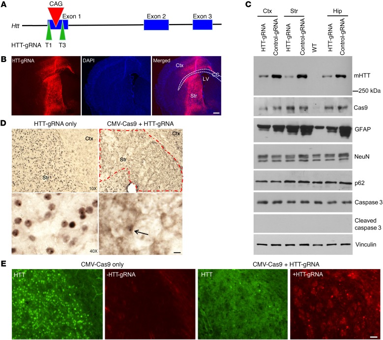
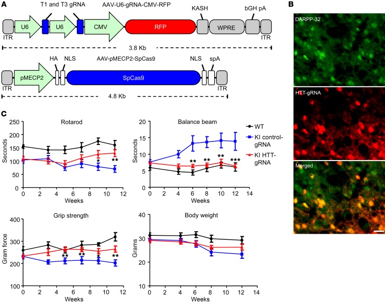
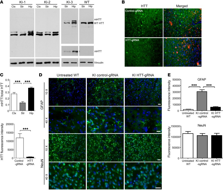
Huntington's disease is a neurodegenerative disorder caused by a polyglutamine repeat in the Huntingtin gene (HTT). Although suppressing the expression of mutant HTT (mHTT) has been explored as a therapeutic strategy to treat Huntington's disease, considerable efforts have gone into developing allele-specific suppression of mHTT expression, given that loss of Htt in mice can lead to embryonic lethality. It remains unknown whether depletion of HTT in the adult brain, regardless of its allele, could be a safe therapy. Here, we report that permanent suppression of endogenous mHTT expression in the striatum of mHTT-expressing mice (HD140Q-knockin mice) using CRISPR/Cas9-mediated inactivation effectively depleted HTT aggregates and attenuated early neuropathology. The reduction of mHTT expression in striatal neuronal cells in adult HD140Q-knockin mice did not affect viability, but alleviated motor deficits. Our studies suggest that non-allele-specific CRISPR/Cas9-mediated gene editing could be used to efficiently and permanently eliminate polyglutamine expansion-mediated neuronal toxicity in the adult brain.


Conflict of interest statement

Conflict of interest: The authors have declared that no conflict of interest exists.

Figures

Figure 1
Figure 1. CRISPR/Cas9 depletes the ubiquitous expression of mHTT in homozygous HD140Q-KI mouse striatum.
(A) Schematics of the designed HTT-gRNA (T1 and T3). (B) Immunofluorescence showing the transduction of AAV-HTT-gRNA in the striatum and part of the cortex. Ctx, cortex; Str, striatum; CC, corpus callosum; LV, lateral ventricle. Scale bar: 100 μm. (C) Different brain regions from 9-month-old homozygous HD140Q-KI mice injected with AAV-CMV-Cas9 and AAV-HTT-gRNA (T1 and T3) or control-gRNA were analyzed by Western blotting with 1C2 for mHTT and antibodies against Cas9, GFAP, NeuN, p62, caspase 3, and cleaved caspase 3. Vinculin was used as a loading control. Hip, hippocampus. (D) Low- and high-magnification images show the reduction of nuclear HTT and HTT aggregates in the AAV-HTT-gRNA/AAV-CMV-Cas9–injected area in 9-month-old homozygous HD140Q-KI mice compared with the contralateral striatum injected with AAV-HTT-gRNA only. Arrow indicates a remaining cell with nuclear HTT inclusion. Scale bar: 10 μm. The red dashed outline indicates the injected region where mHTT aggregates are markedly reduced. (E) Double immunostaining confirmed the depletion of mHTT in the area expressing HTT-gRNA in the injected striatum of 9-month-old homozygous HD140Q-KI mice. The striatum of a HD140Q-KI mouse injected with AAV-CMV-Cas9 only was used as a control. Scale bar: 20 μm.
Figure 2
Figure 2. Behavioral analysis of heterozygous HD140Q-KI mice with depletion of neuronal HTT in the striatum by AAV-HTT-gRNA/AAV-MECP2-Cas9 injection.
(A) Schematics showing the viral vectors used. HA, human influenza hemagglutinin; ITR, inverted terminal repeat; KASH, Klarsicht, ANC-1, Syne Homology; WPRE, woodchuck hepatitis virus post-transcriptional regulatory element. (B) Double immunostaining with anti–DARRP-32 indicated that medium spiny neurons expressed AAV-HTT-gRNA. Scale bar: 10 μm. (C) Motor functions of heterozygous HD140Q-KI mice injected with AAV-HTT-gRNA/AAV-MECP2-Cas9 (KI HTT-gRNA) or AAV-control-gRNA/AAV-MECP2-Cas9 (KI control-gRNA) and WT mice injected with AAV-control-gRNA/AAV-MECP2-Cas9 (WT) were evaluated using rotarod, balance beam, and grip strength tests at different time points after injection (n = 12 for each group; *P < 0.05, **P < 0.012, and ***P < 0.001, by 2-way ANOVA with Bonferroni’s test, comparing the KI HTT-gRNA group with the KI control-gRNA group).Data represent the mean ± SEM.
Figure 3
Figure 3. Removal of mHTT in neuronal cells alleviates neuropathology in 13-month-old heterozygous HD140Q-KI mouse striatum.
(A) Western blotting shows the reduction of mHTT in brain tissues from 3 heterozygous HD140Q-KI (KI-1, KI-2, and KI-3) and WT mice. 2166 Antibody was used to show both mHTT and WT HTT. 1C2 Antibody was used to show only mHTT. Replicate samples run on separated blots are presented. (B) Double immunostaining with 1C2 antibody confirmed the depletion of mHTT by AAV-HTT-gRNA. A heterozygous HD140Q-KI mouse injected with AAV-control-gRNA served as a control. Scale bar: 20 μm. (C) Quantitative assessments of the relative ratio of mHTT to total HTT in A (left; n = 8; ***P < 0.001, by 1-way ANOVA with Tukey’s test) and relative levels of mHTT staining in B (right; n = 8; ***P < 0.001, by Student’s t test). (D) Double immunostaining of striatum (from 9-month-old injected mice examined at 13 months of age) shows decreased GFAP levels by HTT-gRNA compared with control-gRNA. There was no difference in NeuN staining. Scale bars: 20 μm. (E) Quantitative assessment of the relative levels of GFAP and NeuN staining (n = 8). The staining intensity for each mouse was the average from three ×10 images. ***P < 0.001, by 1-way ANOVA with Tukey’s test. Data represent the mean ± SEM.

References

    1. Bates GP, et al. Huntington disease. Nat Rev Dis Primers. 2015;1:15005. - PubMed
    1. Carroll JB, et al. Potent and selective antisense oligonucleotides targeting single-nucleotide polymorphisms in the Huntington disease gene/allele-specific silencing of mutant huntingtin. Mol Ther. 2011;19(12):2178–2185. doi: 10.1038/mt.2011.201. - DOI - PMC - PubMed
    1. Drouet V, et al. Allele-specific silencing of mutant huntingtin in rodent brain and human stem cells. PLoS One. 2014;9(6):e99341. doi: 10.1371/journal.pone.0099341. - DOI - PMC - PubMed
    1. Monteys AM, Ebanks SA, Keiser MS, Davidson BL. CRISPR/Cas9 editing of the mutant huntingtin allele in vitro and in vivo. Mol Ther. 2017;25(1):12–23. doi: 10.1016/j.ymthe.2016.11.010. - DOI - PMC - PubMed
    1. Harper SQ, et al. RNA interference improves motor and neuropathological abnormalities in a Huntington’s disease mouse model. Proc Natl Acad Sci U S A. 2005;102(16):5820–5825. doi: 10.1073/pnas.0501507102. - DOI - PMC - PubMed