. 2017 Jun 30;127(7):2855-2867.
doi: 10.1172/JCI89934. Epub 2017 Jun 19.

Lipogenic transcription factor ChREBP mediates fructose-induced metabolic adaptations to prevent hepatotoxicity

Affiliations

Lipogenic transcription factor ChREBP mediates fructose-induced metabolic adaptations to prevent hepatotoxicity

Deqiang Zhang et al. J Clin Invest. .

Abstract

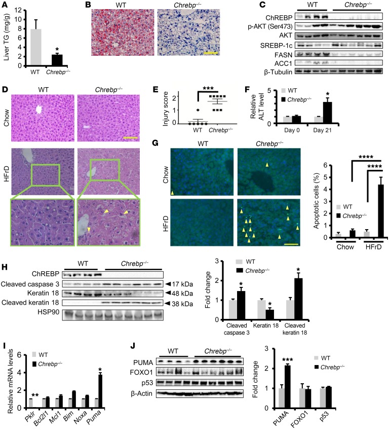
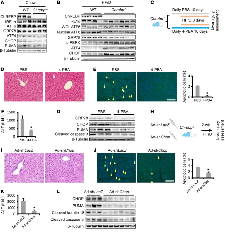
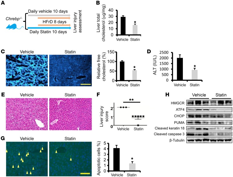
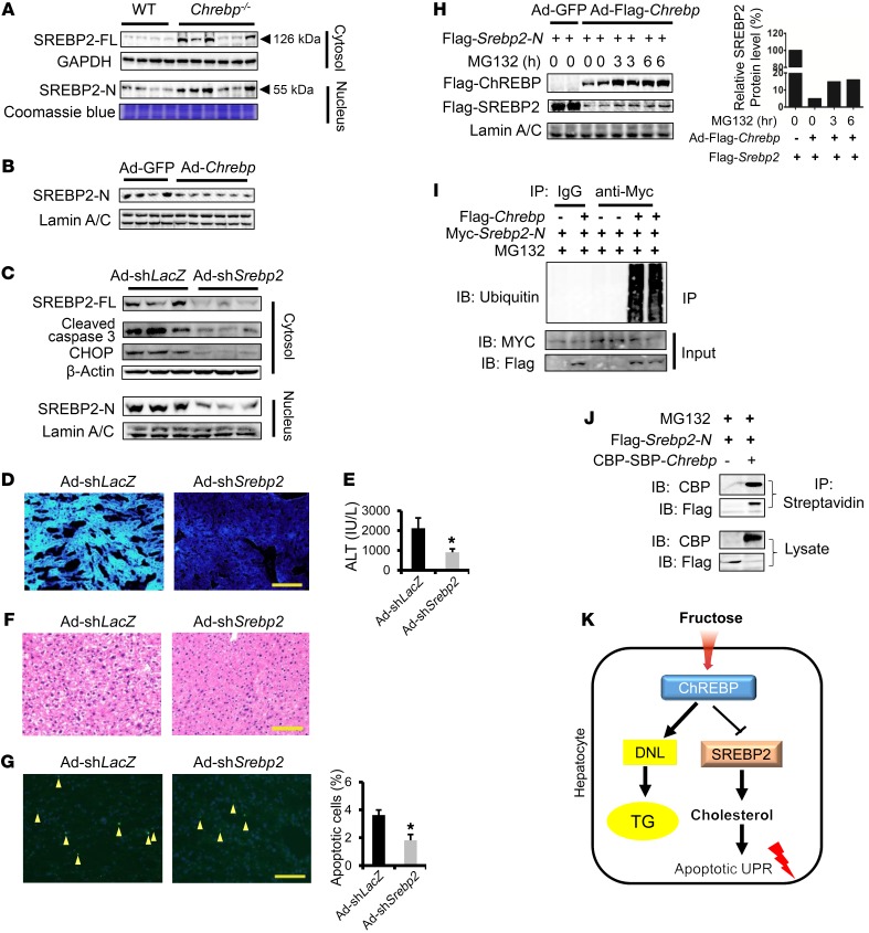
Epidemiologic and animal studies implicate overconsumption of fructose in the development of nonalcoholic fatty liver disease, but the molecular mechanisms underlying fructose-induced chronic liver diseases remain largely unknown. Here, we have presented evidence supporting the essential function of the lipogenic transcription factor carbohydrate response element-binding protein (ChREBP) in mediating adaptive responses to fructose and protecting against fructose-induced hepatotoxicity. In WT mice, a high-fructose diet (HFrD) activated hepatic lipogenesis in a ChREBP-dependent manner; however, in Chrebp-KO mice, a HFrD induced steatohepatitis. In Chrebp-KO mouse livers, a HFrD reduced levels of molecular chaperones and activated the C/EBP homologous protein-dependent (CHOP-dependent) unfolded protein response, whereas administration of a chemical chaperone or Chop shRNA rescued liver injury. Elevated expression levels of cholesterol biosynthesis genes in HFrD-fed Chrebp-KO livers were paralleled by an increased nuclear abundance of sterol regulatory element-binding protein 2 (SREBP2). Atorvastatin-mediated inhibition of hepatic cholesterol biosynthesis or depletion of hepatic Srebp2 reversed fructose-induced liver injury in Chrebp-KO mice. Mechanistically, we determined that ChREBP binds to nuclear SREBP2 to promote its ubiquitination and destabilization in cultured cells. Therefore, our findings demonstrate that ChREBP provides hepatoprotection against a HFrD by preventing overactivation of cholesterol biosynthesis and the subsequent CHOP-mediated, proapoptotic unfolded protein response. Our findings also identified a role for ChREBP in regulating SREBP2-dependent cholesterol metabolism.


Conflict of interest statement

Conflict of interest: The authors have declared that no conflict of interest exists.

Figures

Figure 1
Figure 1. Loss of Chrebp sensitizes mice to HFrD-induced liver injury.
Eight-week-old WT and Chrebp–/– mice were fed a HFrD (70% calories from free fructose) for two weeks (n = 4 for WT, n = 6 for Chrebp–/– mice). (AC) Loss of Chrebp blocked HFrD-induced hepatic lipogenesis. After 2 weeks of HFrD feeding, (A) a liver triglyceride (TG) assay and (B) Oil Red O staining were performed to assess lipid accumulation in the livers of Chrebp–/– mice and their WT littermates. (C) Protein levels of lipogenic enzymes were assessed by Western blotting (protein quantification is shown in Supplemental Figure 4G). (DF) HFrD feeding induced liver injury in Chrebp–/– mice. (D) After H&E staining of livers from Chrebp–/– mice and their WT littermates fed either regular chow or a HFrD, (E) liver injury was scored blindly on a scale of 0 to 2. Mallory-Denk bodies are indicated by yellow arrows. (F) Serum ALT levels were measured at the start and end of HFrD feeding. (G and H) HFrD feeding induced apoptosis in Chrebp–/– mouse livers. Apoptosis was determined by (G) TUNEL staining and (H) Western blotting for apoptotic markers. Apoptotic cells are indicated by arrowheads. (I and J) HFrD feeding increased the expression of PUMA at both (I) mRNA and (J) protein levels. *P < 0.05, **P < 0.01, and ***P < 0.001, by 2-tailed Student’s t test; ****P < 0.0001, by 1-way ANOVA with Tukey’s post-hoc test. Data represent the mean ± SEM. Scale bars: 100 μm; original magnification, ×400 (enlarged images in bottom panels of D).
Figure 2
Figure 2. Restoring Chrebp expression rescues liver injury in HFrD-fed Chrebp–/– mice.
Eight-week-old male Chrebp–/– mice were injected with either Chrebp-expressing adenovirus (Ad-Flag-Chrebp, n = 9) or GFP-expressing control adenovirus (Ad-GFP, n = 7). Three mice from each group were dissected three days after adenovirus injection, and Chrebp overexpression was confirmed by (A) Western blotting with anti-Flag and (B) RT-qPCR for Fasn and Pklr. The remaining mice were fed a 70% HFrD for 2 weeks before dissection. (C) Effects of restoring ChREBP on liver triglyceride levels. Liver injury was assessed by (D) H&E staining, (E) TUNEL staining (apoptotic cells are indicated by arrowheads), (F) ALT assay, and (G) Western blotting for apoptotic markers. *P < 0.05, by 2-tailed Student’s t test. Data represent the mean ± SEM. Scale bars: 100 μm.
Figure 3
Figure 3. HFrD activates the proapoptotic branch of the UPR in Chrebp–/– mouse liver.
(A) Comparable levels of ER stress in the livers of regular chow–fed Chrebp–/– mice and their WT littermates. Livers of 8-week-old male Chrebp–/– mice and their WT littermates (n = 3) on a regular chow diet were subjected to Western blotting for ER stress markers. (B) HFrD feeding activated the proapoptotic branch of the UPR in Chrebp–/– mouse livers. Male and female Chrebp–/– mice and their WT littermates were fed a 70% HFrD for 2 weeks before dissection (n = 4 for WT, n = 6 for Chrebp–/– mice). Protein levels of components of adaptive and proapoptotic branches of ER stress in the liver were assessed with Western blotting (protein level quantification is shown in Supplemental Figure 8). WCL, whole-cell lysate. (CG) Administration of the chemical chaperone 4-BPA protected Chrebp–/– mice from HFrD-induced liver injury. (C) Male 8-week-old Chrebp–/– mice were pretreated with 4-PBA (1 g/kg BW/day) or PBS by oral gavage for 2 days (n = 3/group), followed by 8 days of HFrD feeding plus 4-PBA or PBS gavage. Liver injury was determined by (D) H&E staining, (E) TUNEL staining (apoptotic cells are indicated by arrowheads), (F) ALT assay, and (G) Western blotting for apoptotic markers. (HL) Blocking the proapoptotic branch of the UPR protected Chrebp–/– mice from HFrD-induced liver injury. (H) Male and female 8-week-old Chrebp–/– mice were injected with either Chop-knockdown adenovirus (Ad-shChop, n = 6) or control adenovirus (Ad-shLacZ, n = 3) and then fed a HFrD for 2 weeks. Liver injury was assessed by (I) H&E staining, (J) TUNEL staining (apoptotic cells are indicated by arrowheads; original magnification, ×200), (K) ALT assay, and (L) Western blotting for apoptotic markers. *P < 0.05, by 2-tailed Student’s t test. Data represent the mean ± SEM. Scale bars: 100 μm.
Figure 4
Figure 4. ChREBP protects mice from HFrD-induced liver injury via suppression of cholesterol biosynthesis.
Microarray analysis was performed with RNA samples pooled from the livers of regular chow–fed WT mice (n = 7) versus 70% HFrD–fed WT mice (n = 7) or from the livers of HFrD-fed WT mice (n = 4) versus HFrD-fed Chrebp–/– mice (n = 6). Both male and female mice were used in all 4 groups. (A) List of ChREBP-regulated genes in mouse livers in response to high-fructose feeding (genes involved in de novo cholesterol biosynthesis are highlighted in red). (B) Enrichment of genes in cholesterol biosynthesis in HFrD-fed Chrebp–/– mouse liver by PANTHER pathway analysis. (C and D) Elevation of genes in cholesterol biosynthesis in the livers of HFrD-fed Chrebp–/– mice. Increased levels of cholesterol biosynthesis genes in HFrD-fed Chrebp–/– mouse livers were confirmed by (C) RT-qPCR and (D) Western blotting. (E and F) Restoring hepatic ChREBP expression suppressed cholesterol biosynthesis genes and HMGCR protein in the livers of HFrD-fed Chrebp–/– mice. (G and H) Increased free cholesterol content in Chrebp–/– mouse livers after HFrD feeding. (G) Total cholesterol and (H) free cholesterol levels in the liver were assessed with a cholesterol quantification kit and filipin staining, respectively. (I) Restoration of hepatic ChREBP expression suppressed free cholesterol loading in the livers of HFrD-fed Chrebp–/– mice. *P < 0.05 and **P < 0.01; an unpaired, 2-tailed Student’s t test was used to determine the P values in CE and GI. Data represent the mean ± SEM. Scale bars: 100 μm.
Figure 5
Figure 5. Blocking cholesterol biosynthesis protects Chrebp–/– mice from fructose-induced liver injury.
(A) Male 8-week-old Chrebp–/– mice were pretreated by oral gavage with atorvastatin (20 mg/kg BW/day) or vehicle for 2 days, followed by 8 days of 70% HFrD feeding plus atorvastatin or vehicle gavage (n = 6/group). (B and C) Liver total cholesterol and free cholesterol levels were determined. Liver injury was assessed by (D) ALT assay, (E and F) H&E staining, and (G) TUNEL staining (arrowheads indicate apoptotic cells). (H) HMGCR and apoptotic markers were measured by Western blotting. *P < 0.05 and **P < 0.01, by 2-tailed Student’s t test.
Figure 6
Figure 6. ChREBP blocks HFrD-induced liver injury in part by degrading SREBP2-N to suppress cholesterol biosynthesis.
(A) HFrD feeding induced both SREBP2-FL and SREBP2-N protein expression in the livers of 70% HFrD–fed Chrebp–/– mice. (B) Restoring ChREBP expression lowered SREBP2-N in the nuclear extract from the livers of HFrD-fed Chrebp–/– mice. (CG) Depletion of Srebp2 by shRNA ameliorated HFrD-induced liver injury in Chrebp–/– mice. Chrebp–/– mice (8 weeks of age) were injected with either Srebp2-knockdown adenovirus (Ad-shSrebp2, n = 3) or control adenovirus (Ad-shLacZ, n = 3) and then fed a HFrD for 2 weeks. (C) Srebp2-knockdown efficiency was confirmed by Western blotting, and (D) its targets expression and filipin staining detected free cholesterol. Liver injury was assessed by (C) Western blotting with antibodies against apoptotic markers, (E) a serum ALT assay, (F) liver H&E staining, and (G) TUNEL staining (arrowheads indicate apoptotic cells). (H) ChREBP promoted SREBP2 protein degradation. U2OS cells were transfected with Flag-Srebp2-N and cotransduced with Ad-GFP or Ad-Flag-Chrebp and then treated with 10 μM MG132 for 3 or 6 hours. ChREBP and SREBP2-N expression levels were assessed by Western blotting with anti-Flag antibody. (I) ChREBP promoted SREBP2 protein ubiquitination. 293A cells were transfected with Myc-Srebp2-N and cotransduced with Ad-GFP or Ad-Flag-Chrebp. Polyubiquitinated SREBP2-N was pulled down by denaturing immunoprecipitation with anti-Myc antibody and detected by Western blotting with antiubiquitin. (J) ChREBP interacted with SREBP2. 293A cells were transfected with Flag-Srebp2-N and cotransfected with pNTAP-CBP-SBP-Chrebp or pNTAP empty vector. The lysate was subjected to immunoprecipitation with streptavidin beads and to Western blotting with CBP or Flag antibodies. (K) Working model: high-fructose–induced ChREBP suppresses free cholesterol loading and protects mice from liver injury via the promotion of SREBP2 degradation. Data shown in H, I, and J are representative results of 3 independent experiments. *P < 0.05, by 2-tailed Student’s t test. Data represent the mean ± SEM. Scale bars: 100 μm. IB, immunoblot; IP, immunoprecipitation.

Comment in

  • ChREBP refines the hepatic response to fructose to protect the liver from injury doi: 10.1172/JCI95008

References

    1. Nomura K, Yamanouchi T. The role of fructose-enriched diets in mechanisms of nonalcoholic fatty liver disease. J Nutr Biochem. 2012;23(3):203–208. doi: 10.1016/j.jnutbio.2011.09.006. - DOI - PubMed
    1. Tiniakos DG, Vos MB, Brunt EM. Nonalcoholic fatty liver disease: pathology and pathogenesis. Annu Rev Pathol. 2010;5:145–171. doi: 10.1146/annurev-pathol-121808-102132. - DOI - PubMed
    1. Neuschwander-Tetri BA. Carbohydrate intake and nonalcoholic fatty liver disease. Curr Opin Clin Nutr Metab Care. 2013;16(4):446–452. doi: 10.1097/MCO.0b013e328361c4d1. - DOI - PubMed
    1. Dekker MJ, Su Q, Baker C, Rutledge AC, Adeli K. Fructose: a highly lipogenic nutrient implicated in insulin resistance, hepatic steatosis, and the metabolic syndrome. Am J Physiol Endocrinol Metab. 2010;299(5):E685–E694. doi: 10.1152/ajpendo.00283.2010. - DOI - PubMed
    1. Ren LP, et al. Differing endoplasmic reticulum stress response to excess lipogenesis versus lipid oversupply in relation to hepatic steatosis and insulin resistance. PLoS ONE. 2012;7(2):e30816. doi: 10.1371/journal.pone.0030816. - DOI - PMC - PubMed

MeSH terms