. 2017 Jun 19:2:17096.
doi: 10.1038/nmicrobiol.2017.96.

Toxoplasma depends on lysosomal consumption of autophagosomes for persistent infection

Affiliations

Toxoplasma depends on lysosomal consumption of autophagosomes for persistent infection

Manlio Di Cristina et al. Nat Microbiol. .

Abstract

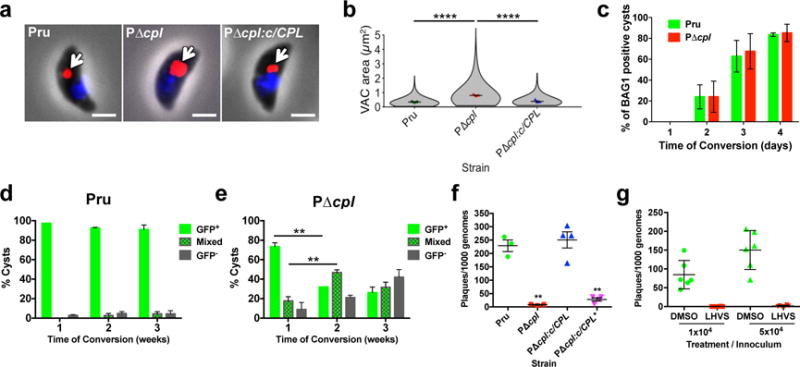
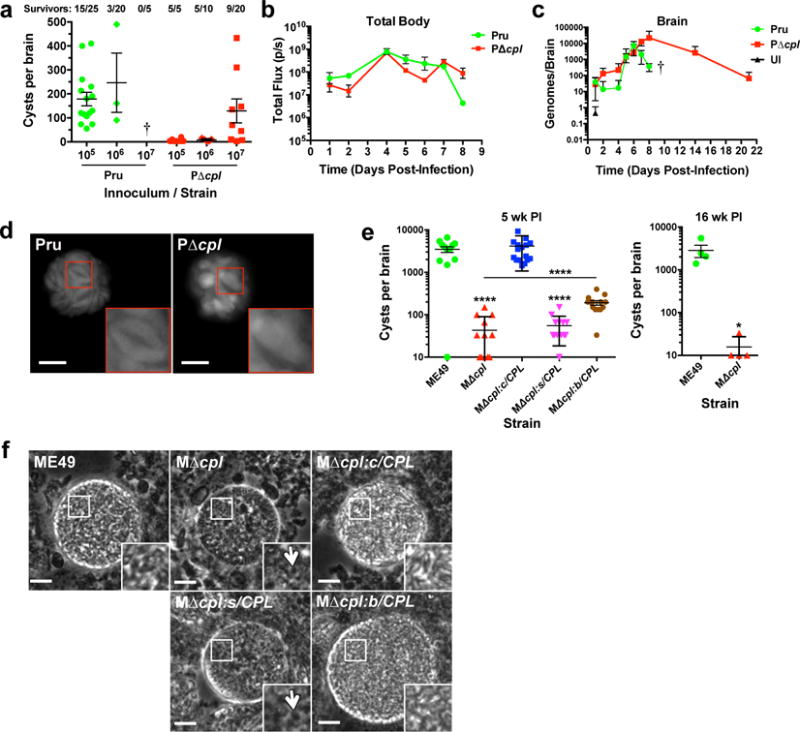
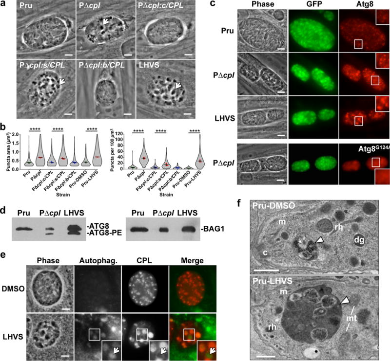
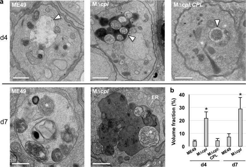
Globally, nearly 2 billion people are infected with the intracellular protozoan Toxoplasma gondii1. This persistent infection can cause severe disease in immunocompromised people and is epidemiologically linked to major mental illnesses2 and cognitive impairment3. There are currently no options for curing this infection. The lack of effective therapeutics is due partly to a poor understanding of the essential pathways that maintain long-term infection. Although it is known that Toxoplasma replicates slowly within intracellular cysts demarcated with a cyst wall, precisely how it sustains itself and remodels organelles in this niche is unknown. Here, we identify a key role for proteolysis within the parasite lysosomal organelle (the vacuolar compartment or VAC) in turnover of autophagosomes and persistence during neural infection. We found that disrupting a VAC-localized cysteine protease compromised VAC digestive function and markedly reduced chronic infection. Death of parasites lacking the VAC protease was preceded by accumulation of undigested autophagosomes in the parasite cytoplasm. These findings suggest an unanticipated function for parasite lysosomal degradation in chronic infection, and identify an intrinsic role for autophagy in the T. gondii parasite and its close relatives. This work also identifies a key element of Toxoplasma persistence and suggests that VAC proteolysis is a prospective target for pharmacological development.


Conflict of interest statement

Author Information

Figures

Figure 1
Figure 1. VAC proteolytic activity is required for Toxoplasma viability and persistence in vitro
a, Extracellular tachyzoites were stained with α-CPB (red) to mark the VAC and DAPI (blue) to identify the nucleus. An arrow indicates the localization of the VAC. Scale bar, 2 μm. b, Violin plots of VAC size. Data are mean ± SEM of 3 biological replicates, with the following number of parasites evaluated: Pru, 317, 153, 154; PΔcpl, 271, 177, 176; PΔcpl:c/CPL, 293, 153, 153. Shape indicates distribution of the pooled data. ****, p<0.0001 Mann Whitney test. c, In vitro differentiation based on expression of the bradyzoite specific antigen BAG1. Cultures were differentiated for the indicated time, fixed, stained with α-BAG1 and enumerated by fluorescence microscopy. Bars are mean ± SEM of 2 biological replicates. Experiment 1 assessed for Pru 205, 214, 204, and 208 parasitophorous vacuoles (PV) on days 1, 2, 3, and 4, respectively; and for PΔcpl, 209, 211, 213, and 202 PV, respectively. Experiment 2 assessed for Pru 212, 202, 208, and 202 PV on days 1, 2,3, and 4, respectively; and for PΔcpl 205, 207, 214, and 208 PV, respectively. Viability of Pru (d) or PΔcpl (e) bradyzoites based on stage specific expression of cytosolic GFP under the LHD2 promoter. Cultures were differentiated, and enumerated by fluorescence microscopy. Green bars indicate cysts that are uniformly GFP positive. Checkerboard bars specify cysts that contain GFP positive and GFP negative bradyzoites. Grey bars indicate cysts that are uniformly GFP negative. Data are mean ± SD of 3 biological replicates each with 100 cysts evaluated. **, p<0.001, two-tailed paired student’s t-test. f, Viability of CPL proficient and deficient strains based on plaquing efficiency. Strains were differentiated for 4 weeks, mechanically liberated from cysts, quantified by qPCR and inoculated onto fresh cell monolayers for plaque formation. PΔcpl:c/CPL* is complemented with catalytically inactive CPL. Bars are mean ± SEM of 3 (Pru, PΔcpl) or 4 (PΔcpl:c/CPL, PΔcpl:c/CPL*) biological replicates each with two technical replicates. **, p<0.01, Mann Whitney test. g, Viability of Pru bradyzoites differentiated for 1 week before treatment with solvent (DMSO) or 1 μM LHVS for 4 weeks. Viability was measure as in (f). n=2 biological replicates each with 3 technical replicates.
Figure 2
Figure 2. VAC proteolytic function is required for Toxoplasma persistence in infected mice
a, Burden of brain cysts in Pru and PruΔcpl infected C57BL/6 mice. Mice were infected with the indicated inoculum for 4 weeks, humanely euthanized and cysts were enumerated microscopically in brain homogenates. Each data point indicates cyst burden in one mouse. Data are mean ± SEM of data pooled from: Pru 105, 4 experiments; Pru 106, 3 experiments; Pru 107, 1 experiment; PΔcpl 105, 1 experiment; PΔcpl 106, 1 experiment; PΔcpl 107, 3 experiments. All of the mice in the Pru 107 group succumbed to the infection during the acute stage, as indicated by a cross. The number of mice that survived 4 weeks is indicated along with the total number of mice in each group. b, Parasite burden throughout the body of infected mice. C57BL/6 mice (4 per group) were infected intraperitoneally with 106 tachyzoites. Total body parasite burden was determined by bioluminescence imaging and measurement of light flux (photons/second) from parasites expressing firefly luciferase. Data are mean ± SEM from one experiment. c, Mice were infected as in panel b and parasite burden in the brain was measured by qPCR. Data are mean ± SEM from one experiment. A cross indicates that Pru infected mice succumbed to the infection prior to imaging on day 10. UI, uninfected. d, Live fluorescence microscopy of cysts in brain homogenates. Brain homogenates were prepared 4 weeks post-infection for fluorescence microscopy of cytosolic GFP in cysts. Insets show enlargements of boxed regions including two Pru bradyzoites and one PΔcpl bradyzoite. Scale bar, 10 μm. e, Burden of brain cysts in mice infected with ME49 WT or transgenic strains. Female CBA/J mice (20 mice per group) were infected intraperitoneally with 500 tachyzoites and humanely euthanized for cyst enumeration 5 weeks (left panel) or 16 weeks (right panel) post-infection. Five week PI data is pooled from two experiments. Sixteen week PI data is from one experiment. MΔcpl:c/CPL is complemented with CPL under its cognate promoter, MΔcpl:s/CPL is complemented with CPL under the SAG1 promoter for expression only in tachyzoites, MΔcpl:b/CPL is complemented with CPL under the BAG1 promoter for expression only in bradyzoites. Data are mean ± SEM. *, p<0.05; ****, p<0.0001, Mann Whitney test comparing MΔcpl to ME49, MΔcpl:s/CPL to ME49, or MΔcpl:b/CPL to MΔcpl (line). f, Phase contrast microscopy of ME49 WT or transgenic strain cysts in brain homogenates. Arrows indicate granular structures in CPL deficient cysts. Scale bar, 10 μm.
Figure 3
Figure 3. CPL deficient bradyzoites develop Atg8-positive autophagosomes associated with the VAC
a, Phase contrast microscopy of in vitro encysted bradyzoites differentiated for 7 days. Arrows indicate cytosolic inclusions in CPL deficient strains. Scale bar, 5 μm. b, Quantification of dark inclusion size (area of individual puncta, left panel) and number (per 100 μm2 of cyst area, right panel) shown as violin plots. Data are mean ± SEM of 3 biological replicates. The following number of total cysts and puncta, respectively, were quantified: Pru 74, 409; PΔcpl 102, 3812; PΔcpl:c/CPL 120, 743; PΔcpl:s/CPL 86, 1324; PΔcpl:b/CPL 101, 464; Pru-DMSO 75, 237; and Pru-LHVS 110, 3024. Shape indicates distribution of the pooled data. ****, p<0.0001 Mann Whitney test. c, Fluorescence microscopy of autophagosomes in bradyzoites expressing tdTomato-Atg8. Bradyzoites were differentiated for 7 days, fixed, and viewed for bradyzoite specific expression of GFP and tdTomato-Atg8. Examples in the top 3 rows are of parasites expressing WT tdTomato-Atg8, whereas the bottom row shows parasites expressing a tdTomato-Atg8 bearing a glycine to alanine mutation rendering it refractory to lipidation. Scale bar, 10 μm. The same scale applies to the fluorescence images, which were captured and processed identically. d, Western blots of bradyzoite lysates probed with α-Atg8 or α-BAG1. e, Fluorescence microscopy of autophagosomes in Pru bradyzoites treated with solvent (DMSO) or 1 μM LHVS. Bradyzoites were differentiated for 7 days, treated for 4 days, stained with cytoID (green), fixed, and stained with α-CPL (red). An arrow indicates an example of CPL at the periphery of an autophagosome shown as an inset of the boxed area. Scale bar, 5 μm. The same scale applies to the fluorescence images. f, Transmission electron microscopy of bradyzoites differentiated for 1 week. Abbreviations used: c, conoid; m, microneme; rh, rhoptry; dg, dense granule; mt, mitochondrion. Scale bars, 500 nm.
Figure 4
Figure 4. CPL deficient bradyzoites develop undigested autophagosomes containing organellar remnants
a, Electron microscopy of intracellular ME49, MΔcpl and MΔcpl:CPL comparing the morphology of VAC (arrowheads) at day 4 showing electron density for this compartment in MΔcpl bradyzoites. High magnification views at day 7 reveal cytosolic material (asterisks) resembling piecemeal of the cytoplasm (ME49) and ER (MΔcpl), suggestive of autophagy activities occurring in the VAC with delayed degradation in the mutant reflected by electron dense luminal content. Scale bars, 500 nm. b. Volume fraction of VAC from representative electron micrographs of parasitophorous vacuoles (PV) from each group (29 and 25 PV for ME49 at d4 and d7, respectively; 30 and 32 PV of MΔcpl at d4 and d7, respectively; and 25 PV for MΔcpl:CPL at d4) showing significant increase of this compartment in parasites lacking CPL compared to control strains. Volume fraction equals the area of the organelle divided by the area of the cell multiplied by 100. Bars indicate mean ± SD. *p<0.005 unpaired student’s t-test.

Comment in

Similar articles

Cited by

References

    1. Montoya JG, Liesenfeld O. Toxoplasmosis. Lancet. 2004;363:1965–1976. - PubMed
    1. Sutterland AL, et al. Beyond the association Toxoplasma gondii in schizophrenia, bipolar disorder, and addiction: systematic review and meta-analysis. Acta Psychiatr Scand. 2015;132:161–179. - PubMed
    1. Bharti AR, et al. Latent Toxoplasma Infection and Higher Toxoplasma gondii Immunoglobulin G Levels Are Associated With Worse Neurocognitive Functioning in HIV-Infected Adults. Clin Infect Dis. 2016 - PMC - PubMed
    1. Parussini F, Coppens I, Shah PP, Diamond SL, Carruthers VB. Cathepsin L occupies a vacuolar compartment and is a protein maturase within the endo/exocytic system of Toxoplasma gondii. Mol Microbiol. 2010;76:1340–1357. - PMC - PubMed
    1. Miranda K, et al. Characterization of a novel organelle in Toxoplasma gondii with similar composition and function to the plant vacuole. Mol Microbiol. 2010;76:1358–1375. - PMC - PubMed