

doi: 10.1038/nmeth.4337.
Epub 2017 Jun 19.
Adaptive optics improves multiphoton super-resolution imaging
Affiliations
- PMID: 28628128
- PMCID: PMC7749710
- DOI: 10.1038/nmeth.4337
Item in Clipboard
Adaptive optics improves multiphoton super-resolution imaging
Nat Methods.
2017 Sep.
Abstract
We improve multiphoton structured illumination microscopy using a nonlinear guide star to determine optical aberrations and a deformable mirror to correct them. We demonstrate our method on bead phantoms, cells in collagen gels, nematode larvae and embryos, Drosophila brain, and zebrafish embryos. Peak intensity is increased (up to 40-fold) and resolution recovered (up to 176 ± 10 nm laterally, 729 ± 39 nm axially) at depths ∼250 μm from the coverslip surface.
Figures
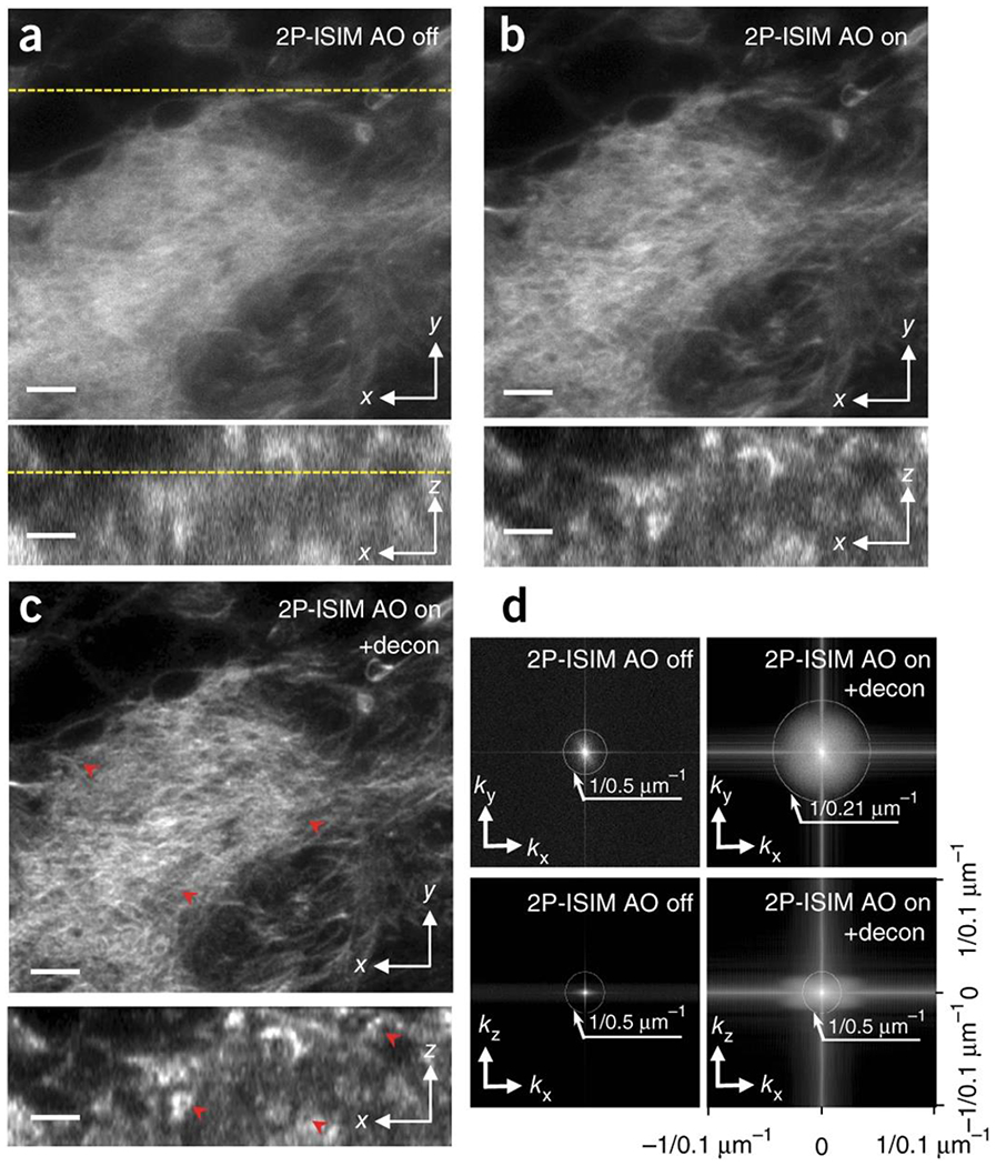
Lateral (top) and axial (bottom) 2P ISIM images of Alexa Fluor 488 phalloidin labeled actin in fixed larval brain lobe, shown without (a) and with (b) adaptive correction, and after subsequent deconvolution (c). The lateral slice is taken 35 μm from the surface of the brain lobe; correspondence with axial slice is indicated with yellow dotted line. Progressive improvements in spatial resolution and contrast are evident in a-c (see also red arrowheads), and further quantified in d), where lateral (top row) and axial (bottom row) MTFs are shown. Note that axial resolution is limited to the step size used when acquiring stacks (0.5 μm for this dataset). See also Supplementary Fig. 6, Supplementary Video 1. Scale bars: 5 μm.
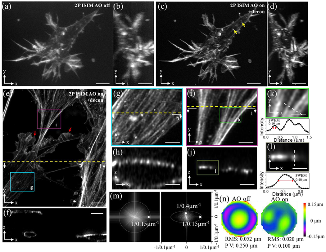
a) Rhodamine-phalloidin stained actin in a primary mouse endothelial cell, embedded in collagen matrix and imaged with 2P ISIM 150 μm from surface of coverslip. Lateral (a) and axial (b) views are shown. c, d) Images as in a, b but after AO correction using a 1 μm fluorescent bead fiducial as a guide-star. Additional cells 100 μm from the coverslip are also shown (lateral view, e) and axial view f). Higher magnification views of boxed regions in e) are shown in g, i along with corresponding axial views h, j. Resolution is estimated from actin bundles, laterally in k and axially in l (higher magnification views of boxed region in i, j respectively), and further quantified in MTFs in m (lateral, left; axial, right). Wavefronts before (left) and after AO correction (right), corresponding to sample in e) are also shown (n). Note that (a-e, g, i) display maximum intensity projections; (f, h, j, l) are single plane cross sections as indicated by dotted lines in (e, g, i) and boxed region in j; and k is a single lateral plane at boxed region indicated in i). Yellow arrows in c) indicate cortical actin structures; red arrows in e) indicate lamellipodial structures. See also Supplementary Video 3. Scale bars: 5 μm (a-f), 2 μm (g-j), 1 μm (k, l).
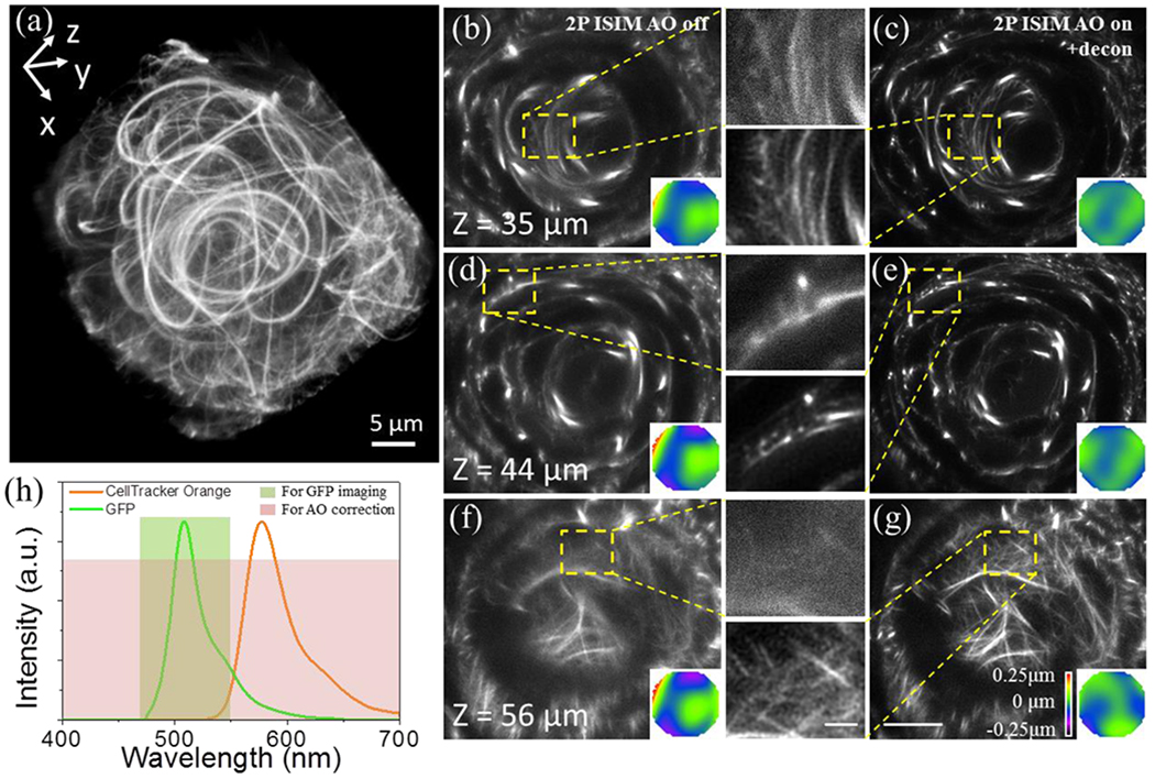
a) Overview rendering of AO-corrected and deconvolved 2P-ISIM volume. Selected slices at indicated axial depth are shown before (b, d, f) and after AO correction and deconvolution (c, e, g), along with wavefront maps (inset) and higher magnification views of yellow dashed rectangular regions. h) Spectra of GFP and CellTracker Orange dye, indicating spectral regions used for AO correction and imaging. See also Supplementary Fig. 10, Supplementary Video 4. Scale bars in b-g: 10 μm (zoomed out views), 2 μm (insets).
References
Publication types
MeSH terms
Grants and funding
LinkOut - more resources
Full Text Sources
Other Literature Sources
Molecular Biology Databases
