. 2017 Aug;14(8):805-810.
doi: 10.1038/nmeth.4335. Epub 2017 Jun 19.

High-speed fixed-target serial virus crystallography

Affiliations

High-speed fixed-target serial virus crystallography

Philip Roedig et al. Nat Methods. 2017 Aug.

Abstract

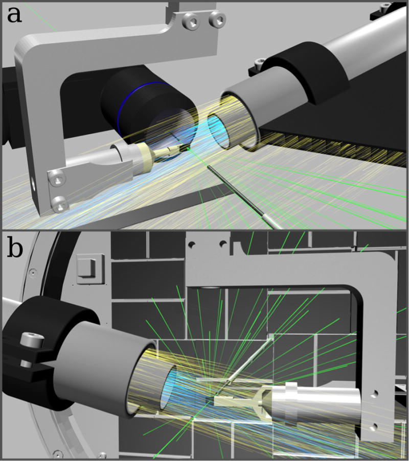
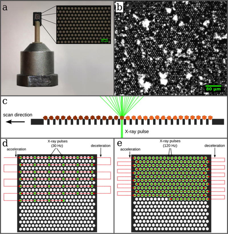
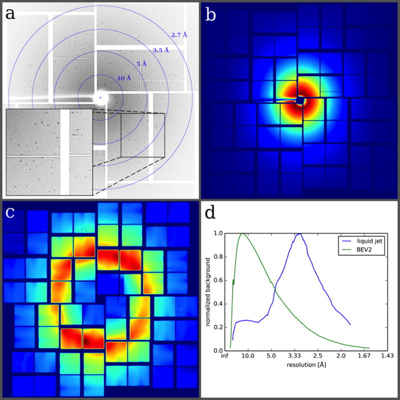
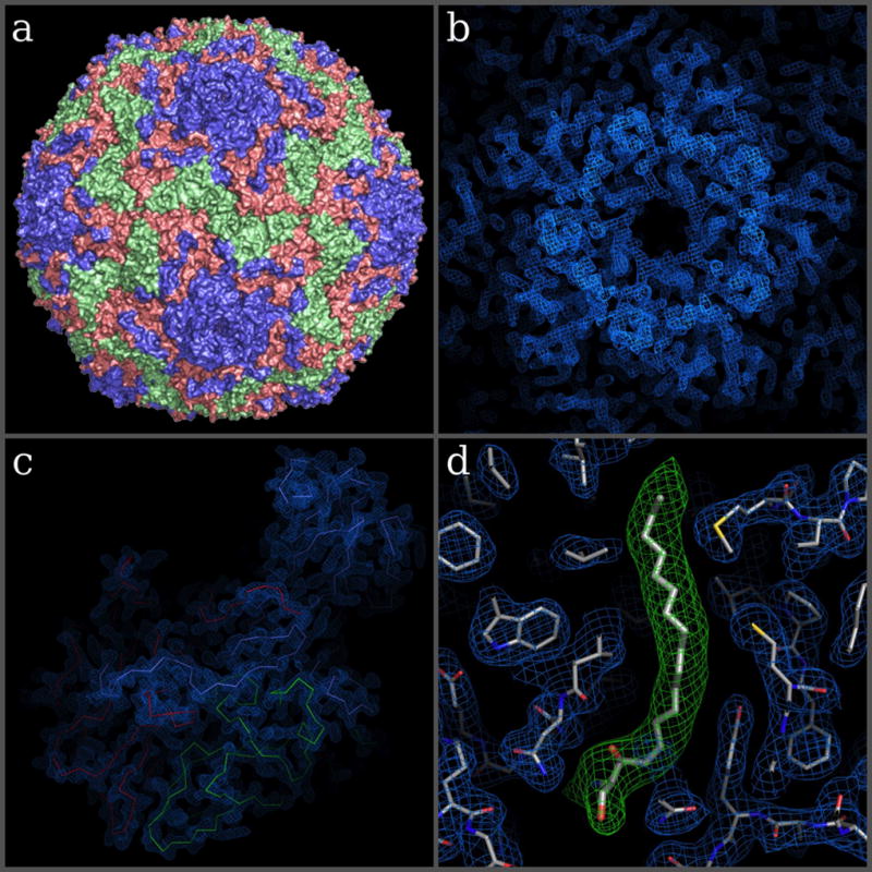
We report a method for serial X-ray crystallography at X-ray free-electron lasers (XFELs), which allows for full use of the current 120-Hz repetition rate of the Linear Coherent Light Source (LCLS). Using a micropatterned silicon chip in combination with the high-speed Roadrunner goniometer for sample delivery, we were able to determine the crystal structures of the picornavirus bovine enterovirus 2 (BEV2) and the cytoplasmic polyhedrosis virus type 18 polyhedrin, with total data collection times of less than 14 and 10 min, respectively. Our method requires only micrograms of sample and should therefore broaden the applicability of serial femtosecond crystallography to challenging projects for which only limited sample amounts are available. By synchronizing the sample exchange to the XFEL repetition rate, our method allows for most efficient use of the limited beam time available at XFELs and should enable a substantial increase in sample throughput at these facilities.


Conflict of interest statement

Competing Financial Interests Statement

A. Meents is one of the CEOs and shareholder of the DESY spin-off company Suna-Precision GmbH. T. Pakendorf and B. Reime are further shareholders. Suna-Precision sells technical equipment for experiments with X-rays including different micro-structured silicon chips for serial crystallography experiments. I. Vartiainen is shareholder and CTO of the company FinnLitho, which produced the silicon chips used for the experiment.

Figures

Figure 1
Figure 1. Low background experimental setup for fast fixed-target SFX experiments using the Roadrunner goniometer
(a) Front view: The silicon chip is raster scanned through the X-ray beam (green) while maintained in a continuous stream of humidified air (blue). A helium sheath flow (yellow) is used to confine the humidity stream and to reduce air scattering. Air scattering is further reduced by helium injection along the beam path. An inline microscope is used for proper chip alignment and definition of the scanning grid. (b) Back view: X-ray diffraction caused by the sample crystals is recorded with a Cornell-SLAC hybrid pixel array detector (CSPAD). After hitting the sample, the primary beam is enclosed by a molybdenum tubule and additional steel tubules, which further absorb air-scattered photons. In b, the inline microscope is not shown for clarity.
Figure 2
Figure 2. Design of the micro-patterned silicon chip and data collection strategy
(a) The chip is attached to a plastic rod for the purpose of thermal isolation. The membrane part within the outer frame consists of micropores with diameters of typically 4 µm–8 µm, which are arranged in a triangular grid (a, inset). (b) The chip acts as a sample holder for more than 20,000 microcrystals, which largely organize themselves according to the pore pattern. (c) After loading, the microcrystals are scanned through the X-ray beam. By shooting through the micropores in the chips the interaction of the X-rays with any support material is further minimized. (d, e) Scanning strategies for measurements performed at room temperature and cryogenic temperatures, respectively (see text for details).
Figure 3
Figure 3. Exemplary BEV2 diffraction pattern and comparison of background scattering levels achievable with different sample delivery methods
(a) Diffraction image of BEV2 microcrystals obtained at the XPP instrument at LCLS using the micro-patterned silicon chip as a sample holder. (b) Due to the efficient removal of successive mother liquor during sample loading, no water ring is observed in the averaged background image of the chip. (c) For comparison, an averaged background image from a typical SFX liquid jet experiment with CPV 17 crystals is shown. (d) The azimuthally averaged radial distribution of both images is plotted as a function of resolution. Both curves are normalized since measurements were performed under different experimental conditions and therefore a direct comparison was not possible.
Figure 4
Figure 4. Overall structure of BEV2 and corresponding high-resolution electron density maps
(a) Surface representation of BEV2 particle as viewed towards an icosahedral 2-fold axis. VP1, VP2 and VP3 are shown in blue, green and red, respectively. (b–c) Electron density maps after one cycle of 5-fold real space averaging using the phases calculated from the current refined model showing the electron density around the 5-fold in b and for a biological protomer in c. (c) C-alpha traces of VP1-3, colored as in a. (d) A close-up view of the electron density for protein residues around the pocket factor binding site of VP1 (blue mesh and thinner sticks) and density for the pocket factor (thicker sticks show a sphingosine fitted to the density, while the green density is for a simulated annealing omit map).

References

    1. Zocher G, et al. A Sialic Acid Binding Site in a Human Picornavirus. PLoS Pathog. 2014;10:e1004401. - PMC - PubMed
    1. Fry EE, Abrescia NGA, Stuart DI. In: Macromolecular Crystallography: conventional and high-throughput methods. Sanderson MR, Skelly JV, editors. 2007. pp. 245–264.
    1. Fry EE, Grimes J, Stuart DI. Virus crystallography. Mol. Biotechnol. 1999;12:13–23. - PubMed
    1. Axford D, et al. In situ macromolecular crystallography using microbeams. Acta Crystallogr. Sect. D Biol. Crystallogr. 2012;68:592–600. - PMC - PubMed
    1. Hope H. Cryocrystallography of biological macromolecules: a generally applicable method. Acta Crystallogr. Sect. B Struct. Sci. Cryst. Eng. Mater. 1988;44:22–26. - PubMed

Methods References

    1. Roedig P, et al. Sample Preparation and Data Collection for High-Speed Fixed-Target Serial Femtosecond Crystallography. Protocol Exchange. 2017 doi: 10.1038/protex.2017.059. - DOI
    1. Walter TS, et al. A procedure for setting up high-throughput nanolitre crystallization experiments. Crystallization workflow for initial screening, automated storage, imaging and optimization. Acta Crystallogr. Sect. D Biol. Crystallogr. 2005;61:651–657. - PMC - PubMed
    1. Wheeler MJ, Russi S, Bowler MG, Bowler MW. Measurement of the equilibrium relative humidity for common precipitant concentrations: Facilitating controlled dehydration experiments. Acta Crystallogr. Sect. F Struct. Biol. Cryst. Commun. 2012;68:111–114. - PMC - PubMed
    1. Ginn HM, Evans G, Sauter NK, Stuart DI. On the release of cppxfel for processing X-ray free-electron laser images. J. Appl. Crystallogr. 2016;49:1065–1072. - PMC - PubMed
    1. Ginn HM, et al. TakeTwo : an indexing algorithm suited to still images with known crystal parameters. Acta Crystallogr. Sect. D Biol. Crystallogr. 2016;72:956–965. - PMC - PubMed

MeSH terms