. 2017 Jun 19:2:17098.
doi: 10.1038/nmicrobiol.2017.98.

HBV RNA pre-genome encodes specific motifs that mediate interactions with the viral core protein that promote nucleocapsid assembly

Affiliations

HBV RNA pre-genome encodes specific motifs that mediate interactions with the viral core protein that promote nucleocapsid assembly

Nikesh Patel et al. Nat Microbiol. .

Abstract

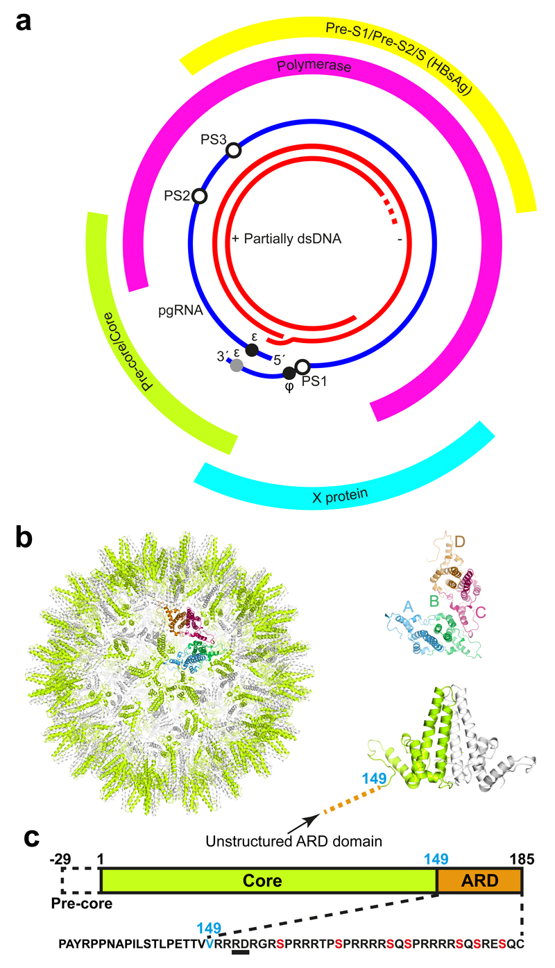
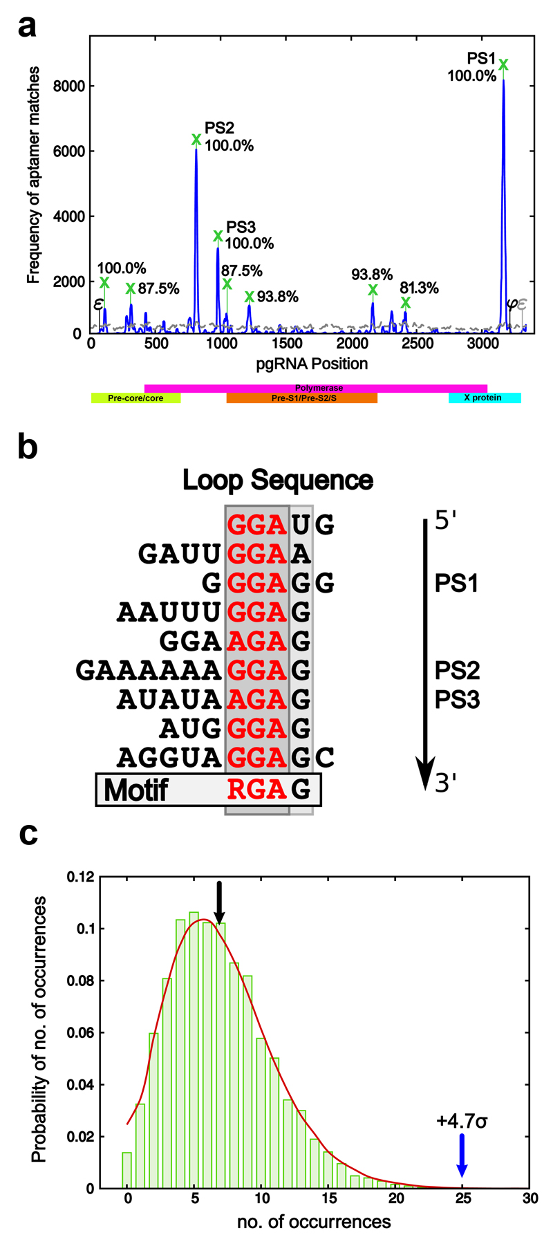
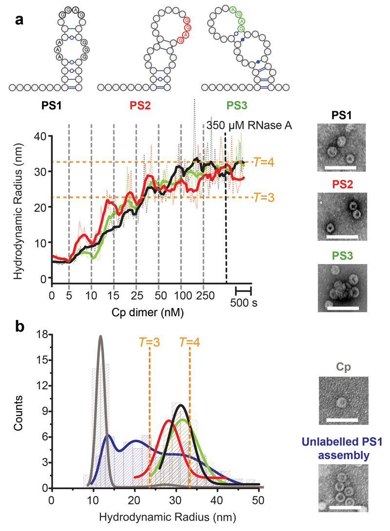
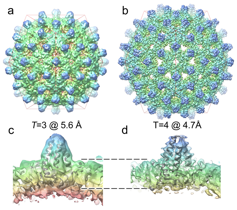
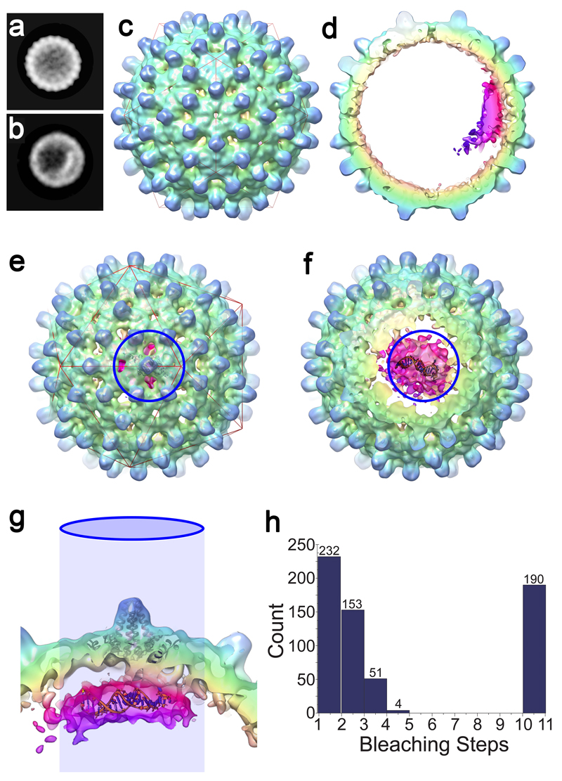
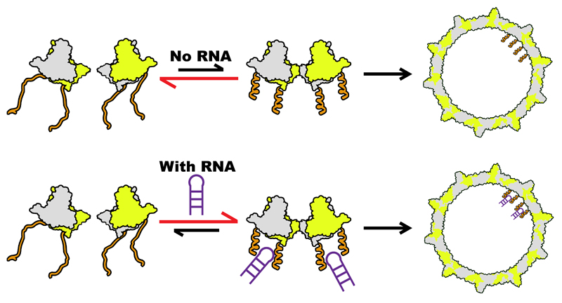
Formation of the hepatitis B virus nucleocapsid is an essential step in the viral lifecycle, but its assembly is not fully understood. We report the discovery of sequence-specific interactions between the viral pre-genome and the hepatitis B core protein that play roles in defining the nucleocapsid assembly pathway. Using RNA SELEX and bioinformatics, we identified multiple regions in the pre-genomic RNA with high affinity for core protein dimers. These RNAs form stem-loops with a conserved loop motif that trigger sequence-specific assembly of virus-like particles (VLPs) at much higher fidelity and yield than in the absence of RNA. The RNA oligos do not interact with preformed RNA-free VLPs, so their effects must occur during particle assembly. Asymmetric cryo-electron microscopy reconstruction of the T = 4 VLPs assembled in the presence of one of the RNAs reveals a unique internal feature connected to the main core protein shell via lobes of density. Biophysical assays suggest that this is a complex involving several RNA oligos interacting with the C-terminal arginine-rich domains of core protein. These core protein-RNA contacts may play one or more roles in regulating the organization of the pre-genome during nucleocapsid assembly, facilitating subsequent reverse transcription and acting as a nucleation complex for nucleocapsid assembly.


Conflict of interest statement

Competing financial interest

AZ is a co-founder and consultant of Assembly BioSciences.

Research in the Zlotnick lab is supported by the NIH and Assembly.

No Assembly BioSciences employee contributed to Dr. Zlotnick’s contribution to this work.

Figures

Figure 1
Figure 1. The Hepatitis B Virus.
(a) The genetic map of HBV showing the partially dsDNA genome and the four open reading frames of the virally encoded proteins: Pre-core/core (Cp), which forms the nucleocapsid (NC) shell; Pre S1/PreS2/S, the envelope embedded HBV antigen (HbsAg); X, which plays a role in numerous aspects of the HBV life-cycle within the cell; the polymerase (P), and the pgRNA with the positions of the 5′ ε, the redundant 3′ ε (grey circle), ϕ and the preferred sites (PSs) studied here highlighted by circles. (b) The HBV NC (left) comprises either 90 (T=3) or 120 Cp dimers (T=4 shown). Cp dimers form characteristic four-helix bundles, two from each monomer, that appear as spikes on the surface (right bottom). The two conformers of the HBV Cp dimer (A/B & C/D) that are needed to create the T=4 particle are also shown (right top). The HBV capsid and protein dimer were obtained from PDB (3J2V). (c) The Cp of the isolate used here is 185 amino acids long (RD dipeptide insertion underlined), with an alpha-helical rich region (149 amino acids long), and a C-terminal ARD. The 149th amino acid, V, is labelled blue for clarity. ARD is rich in both basic amino acids and serines. The latter, highlighted in red, are known sites for phosphorylation, which are thought to play roles in NC assembly.
Figure 2
Figure 2. Identification of conserved PS motifs in the pgRNA.
(a) Matches between aptamers from the HBV core selected library and the reference strain (NC_003977.1) with a Bernoulli score of at least 12 (i.e. all non-contiguous alignments with at least the same probability as a contiguous matching alignment of 12 nucleotides) are shown as a frequency plot (solid blue line). The equivalent frequency plot for the naïve library, i.e. the library before selection has taken place, is shown for comparison (grey dashed line). Peaks occurring in at least 80% of the tested strains are marked by a green cross, with conservation levels indicated as percentages. The peaks with the highest frequency and level of conservation are labelled PS1, PS2 & PS3. (b) Alignment of the loop sequences of representative stem-loops in regions of the genome overlapping with the nine conserved Bernoulli peaks reveals a conserved RGAG motif. (c) Probability distribution showing the proportion of sequences containing a given number of stem-loops with an RGAG containing loop across 10,000 randomised versions of genome. The green bars correspond to such randomised versions of the reference strain, whilst the red line gives corresponding probabilities across all five strains marked by an asterisk in Methods. The black arrow indicates the average number of occurrences over all randomised versions of the reference strain (= 6.85), whilst the blue arrow points to the number of occurrences in the reference strain (= 25), a 4.68 standard deviation from the average. The other tested strains exhibit similar levels of occurrence.
Figure 3
Figure 3. PSs trigger sequence-specific VLP assembly.
(a) Dye end-labelled RNA oligos encompassing PS1 (black), PS2 (red) or PS3 (green) were each assessed for their ability to bind Cp and form VLPs at nanomolar concentrations using smFCS. All reactions contained 15 nM of RNA dye-labelled as described in Methods. Vertical dotted lines indicate points where Cp was added with the final concentrations shown in nM. Samples were allowed to equilibrate between additions. The faint trace represents real time, raw signal, while the thick line represents smoothed data. EM images were recorded of the samples prior to RNase A addition (right). Scale bars represent 100 nm. (b) Hydrodynamic radial distributions of the reactions in (a) were taken following the last addition of Cp (here and throughout). The amount of Cp assembling beyond dimer in the absence and presence of RNA (unlabelled) was compared. At the end of these reactions, Cp was labelled with Alexa Fluor-488 (Methods) and the resulting Rh distributions quantitated for the Cp only (grey) and Cp plus unlabelled PS1 (blue) scenarios. Note, dye-labelling of the Cp dimer prevents it from assembling, implying that this is an end-point measurement. A sample of each was taken for analysis by TEM. smFCS and TEM were repeated in triplicate.
Figure 4
Figure 4. The structures of T=3 and T=4 HBV VLPs suggest a mechanism for the specification of their quasi-conformations.
The icosahedrally-averaged cryo-EM structures of (a) T=3 and (b) T=4 HBV VLPs at 5.6 Å and 4.7 Å resolution, respectively. A red icosahedron is included to assist interpretation of the two reconstructions, which are shown in the same orientation. (c & d) show ˜30 Å thick slabs through the structure of each particle, with a fitted Cp-dimer in each case. The T=3 shell is thicker, indicating that density corresponding to the ARDs is resolved in the T=3, but not the T=4, structure. Rendering both structures at equivalent resolution does not change this interpretation (Supplementary Fig. 5).
Figure 5
Figure 5. Asymmetric RNA feature in T=4 HBV VLPs.
(a & b) 2D views of 42,411 T=4 particles were calculated by maximum-likelihood-based classification in RELION. An asymmetric RNA feature is visible in a subset of these particles (b). (c) An asymmetric 3D reconstruction at 11.5 Å resolution of 10,851 particles containing the asymmetric feature. The asymmetric density for the protein shell is icosahedral, despite the lack of any symmetry averaging. (d) An approximately 40 Å thick slab through the asymmetric HBV VLP reconstruction shows the asymmetric feature bound to one region of the Cp shell, revealing density ascribed to RNA and ARDs within the protein shell (bright cerise, magenta and purple). The figures were rendered in a radial colour scheme (Blue=165Å; Cyan=152Å; Green=139Å; Yellow=126Å; Pink=113Å) using USCF Chimera. (e) The asymmetric RNA density is centred beneath a Cp dimer surrounding one of the 5-fold vertices of the T=4 particle (indicated by the blue circle). A single Cp dimer is fitted as a ribbon diagram into the appropriate position using the ‘Fit in map’ function in UCSF Chimera. (f) As the front of the map is slabbed away, the density within is revealed. Shown and manually fitted is a single copy of PS1 as a ribbon diagram (modelled in RNA Composer). (g) Side-view of the same portion of the map, with the view oriented by the projected blue circle. Discrete fingers of density are visible between the Cp layer and RNA density, which is large enough to accommodate 2-4 RNA oligonucleotides. (h) Histogram of photobleaching steps from 630 individual fluorescent spots on a grid containing PS1 HBV VLPs. Spots containing >10 steps resulted from traces exhibiting exponential decay, which were assumed to be aggregates in which multiple bleaching steps occur simultaneously. Photobleaching was performed in duplicate.
Figure 6
Figure 6. Proposed model of HBV NC assembly.
ARD (orange) within a Cp dimer (green and grey) inhibit formation of a dimer of dimers, the first intermediate on the pathway to NC assembly. Reducing the net charge on the ARD by phosphorylation or PS RNA (purple, bottom) binding allows this structure to form more easily, triggering NC formation. At concentrations higher than those mimicking in vivo conditions as used here, the unmodified dimer of dimers forms and particles self-assemble without RNA or will bind RNA non-specifically to produce the same outcome.

Comment in

  • Viral infection: Packing to leave.
    York A. York A. Nat Rev Microbiol. 2017 Aug;15(8):450-451. doi: 10.1038/nrmicro.2017.81. Epub 2017 Jul 10. Nat Rev Microbiol. 2017. PMID: 28690322 No abstract available.

References

    1. WHO. Weekly epidemiological record. WHO. 2009;84:405–420.
    1. Tillmann HL. Antiviral therapy and resistance with hepatitis B virus infection. World Journal of Gastroenterology. 2007;13:125–140. - PMC - PubMed
    1. Murray K, et al. Protective immunisation against hepatitis B with an internal antigen of the virus. J Med Virol. 1987;23:101–107. - PubMed
    1. Nassal M. Hepatitis B viruses: reverse transcription a different way. Virus Res. 2008;134:235–249. - PubMed
    1. Seeger C, Zoulim F, Mason WS. Hepadnaviruses. Vol. 2. Fields Virology; 2007. pp. 2977–3209.