Review
. 2017 Oct;152(2):185-194.
doi: 10.1111/imm.12778. Epub 2017 Jul 17.

Signals that drive T follicular helper cell formation

Affiliations
Review

Signals that drive T follicular helper cell formation

Louise M C Webb et al. Immunology. 2017 Oct.

Abstract

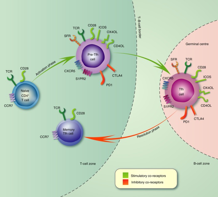
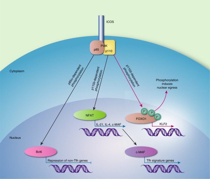
T follicular helper (Tfh) cells are a distinct type of CD4+ T cell specialized in providing help to B cells during the germinal centre (GC) reaction. As such, they are critical determinants of the quality of an antibody response following antigen challenge. Excessive production of Tfh cells can result in autoimmunity whereas too few can result in inadequate protection from infection. Hence, their differentiation and maintenance must be tightly regulated to ensure appropriate but limited help to B cells. Unlike the majority of other CD4+ T-cell subsets, Tfh cell differentiation occurs in three phases defined by their anatomical location. During each phase of differentiation the emerging Tfh cells express distinct patterns of co-receptors, which work together with the T-cell receptor (TCR) to drive Tfh differentiation. These signals provided by both TCR and co-receptors during Tfh differentiation alter proliferation, survival, metabolism, cytokine production and transcription factor expression. This review will discuss how engagement of TCR and co-receptors work together to shape the formation and function of Tfh cells.

Keywords: T follicular helper cell; activation; co-stimulation; inhibitory/activating receptors; signal transduction.


Figures

Figure 1
Figure 1
Expression of co‐receptors during the three phases of T follicular helper (Tfh) cell differentiation. Schematic depicting the multistage and location‐specific nature of Tfh cell differentiation. The expression of co‐receptors is shown at each stage with stimulatory receptors coloured green and inhibitory receptors coloured red. During the first phase of differentiation (days 0–3) naive CD4+ T cells are activated by dendritic cells in the T‐cell zone where they proliferate and alter expression of co‐receptors. This allows the resulting pre‐Tfh cell to migrate towards the T–B‐cell border where it engages with antigen‐specific B cells. Co‐receptors expressed by Tfh cells also modulate the signals transduced through the T‐cell receptor, allowing their differentiation. In the second phase (days 4–5) antigen is presented by B cells and the co‐receptors expressed by pre‐Tfh cells are used symbiotically to both induce Tfh cell differentiation and provide co‐stimulation to B cells. In the third phase (days 6–10), Tfh cells engage with germinal centre (GC) B cells within the GC. Tfh cells provide a limiting source of help to ensure that appropriate B cells differentiate into antibody‐secreting cells and memory B cells. During the resolution phase the Tfh cell can leave the GC and in the absence of further antigenic stimulation resides as a long‐lived memory Tfh cell in the periphery.
Figure 2
Figure 2
The roles of phosphatidlyinositol 3‐kinase (PI3K) subunits in inducible T‐cell co‐stimulator (ICOS) ‐mediated T follicular helper (Tfh) cell differentiation. The regulatory p85α subunit forms a complex with osteopontin (OPN). This complex migrates to the nucleus to protect B‐cell lymphoma 6 (Bcl6) from degradation. Signals through the catalytic p110δ subunit are responsible for nuclear factor of activated T cell (NFAT) activation allowing transcription of interleukin‐21 (IL‐21), IL‐4 and c‐MAF. The transcription factor c‐MAF drives the expression of Tfh cell genes. PI3K also induces phosphorylation of FOXO1, promoting its nuclear egress and so preventing it from activating the transcription factor KLF2, which inhibits Tfh cell differentiation.

References

    1. Suan D, Sundling C, Brink R. Plasma cell and memory B cell differentiation from the germinal center. Curr Opin Immunol 2017; 45:97–102. - PubMed
    1. Bannard O, Cyster JG. Germinal centers: programmed for affinity maturation and antibody diversification. Curr Opin Immunol 2017; 45:21–30. - PubMed
    1. Crotty S. T follicular helper cell differentiation, function, and roles in disease. Immunity 2014; 41:529–42. - PMC - PubMed
    1. Vinuesa CG, Linterman MA, Yu D, MacLennan IC. Follicular helper T cells. Annu Rev Immunol 2016; 34:335–68. - PubMed
    1. Read KA, Powell MD, Oestreich KJ. T follicular helper cell programming by cytokine‐mediated events. Immunology 2016; 149:253–61. - PMC - PubMed

Publication types

LinkOut - more resources