

Real-time imaging of epileptic seizures in rats using electrical impedance tomography
- PMID: 28628556
- PMCID: PMC5491225
- DOI: 10.1097/WNR.0000000000000823
Real-time imaging of epileptic seizures in rats using electrical impedance tomography
Abstract
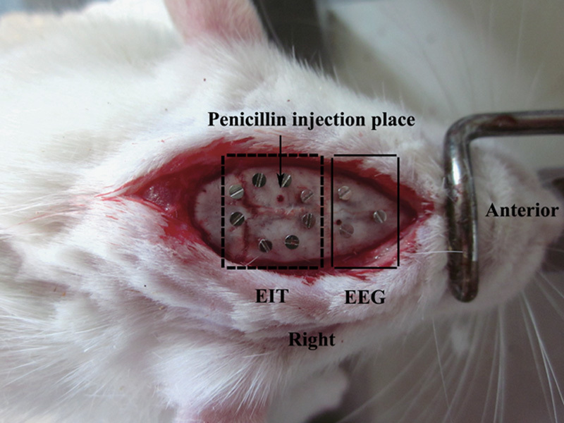
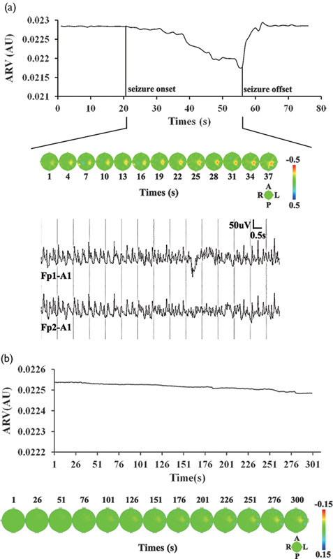
The presence of multiple or diffuse lesions on imaging is a contraindication to surgery for patients with intractable epilepsy. Theoretically, as a functional imaging technique, electrical impedance tomography (EIT) can accurately image epileptic foci. However, most current studies are limited to examining epileptic spikes and few studies use EIT for real-time imaging of seizure activity. Moreover, little is known about changes in electrical impedance during seizures. In this study, we used EIT to monitor seizure progression in real time and analyzed changes in electrical impedance during seizures. EIT and electroencephalography data were recorded simultaneously in rats. Sixty-three seizures were recorded from the cortices of eight rats. During 54 seizures, the average impedance decreased by between 4.86 and 9.17% compared with the baseline. Compared with the control group, the average impedance of the experimental group decreased significantly (P=0.004). Our results indicate that EIT can be used to detect and image electrical impedance reduction within lesions during epileptic seizures.
Figures

References
-
- Spencer S, Huh L. Outcomes of epilepsy surgery in adults and children. Lancet Neurol 2008; 7:525–537. - PubMed
-
- O’Brien TJ, So EL, Mullan BP, Hauser MF, Brinkmann BH, Bohnen NI, et al. Subtraction ictal SPECT co-registered to MRI improves clinical usefulness of SPECT in localizing the surgical seizure focus. Neurology 1998; 50:445–454. - PubMed
-
- la Fougère C, Rominger A, Förster S, Geisler J, Bartenstein P. PET and SPECT in epilepsy: a critical review. Epilepsy Behav 2009; 15:50–55. - PubMed
-
- Xu CH, Wang L, Shi XT, You FS, Fu F, Liu RG, et al. Real-time imaging and detection of intracranial haemorrhage by electrical impedance tomography in a piglet model. J Int Med Res 2010; 38:1596–1604. - PubMed
-
- Abascal JF, Arridge SR, Bayford RH, Holder DS. Comparison of methods for optimal choice of the regularization parameter for linear electrical impedance tomography of brain function. Physiol Meas 2008; 29:1319–1334. - PubMed
MeSH terms
LinkOut - more resources
Full Text Sources
Other Literature Sources
Medical
