. 2017 Jun 19;13(6):e1006454.
doi: 10.1371/journal.ppat.1006454. eCollection 2017 Jun.

The human cytomegalovirus glycoprotein pUL11 acts via CD45 to induce T cell IL-10 secretion

Affiliations

The human cytomegalovirus glycoprotein pUL11 acts via CD45 to induce T cell IL-10 secretion

Jasmin Zischke et al. PLoS Pathog. .

Abstract

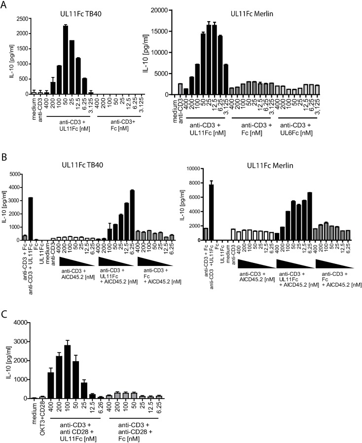
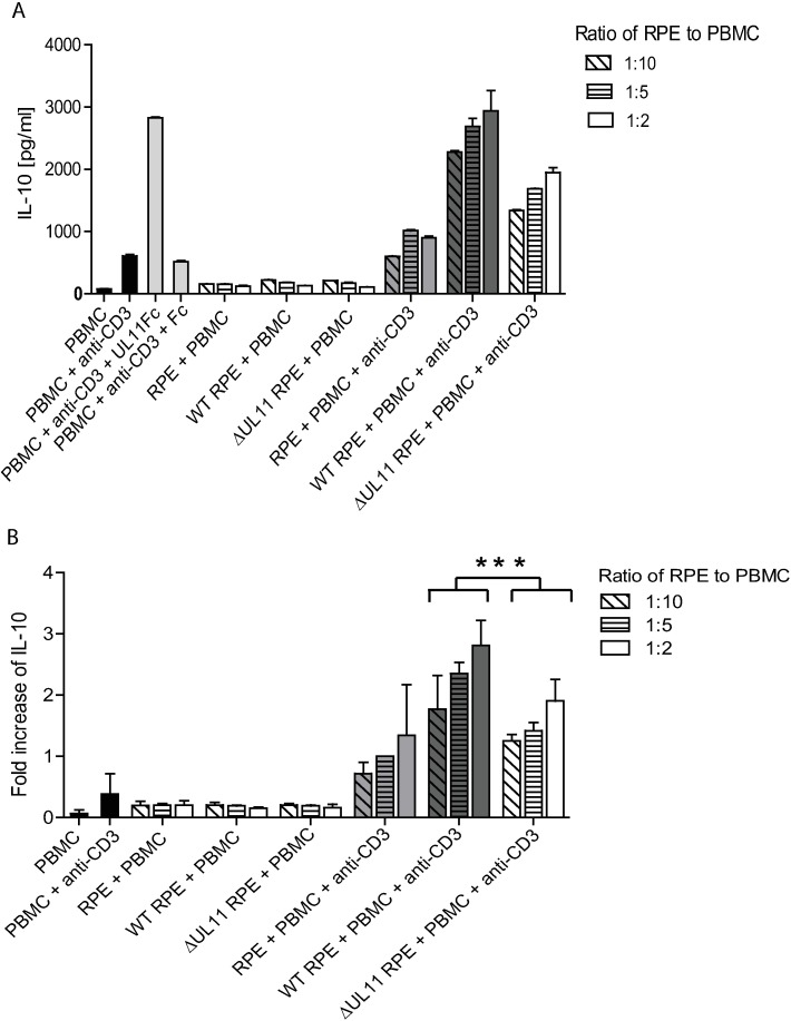
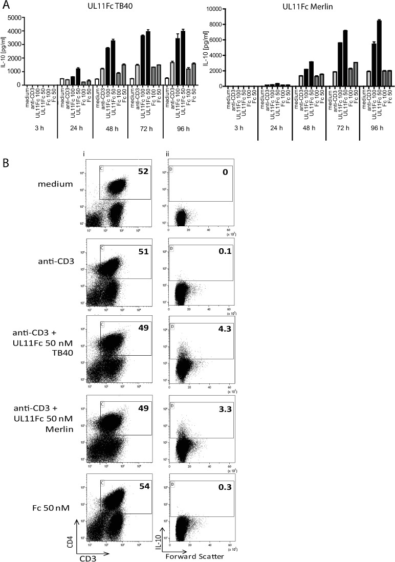
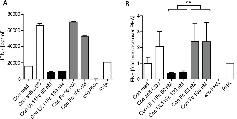
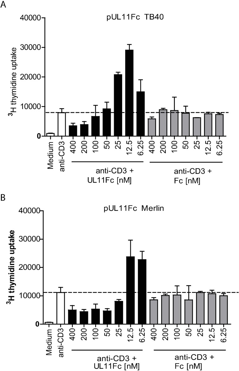
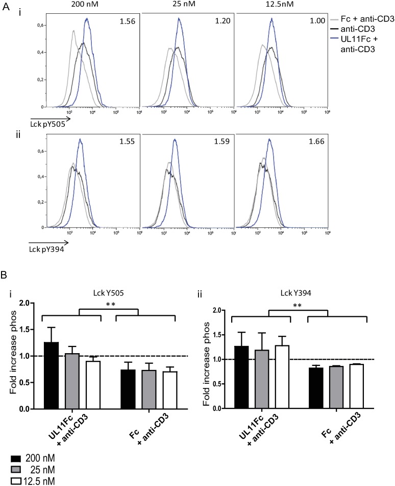
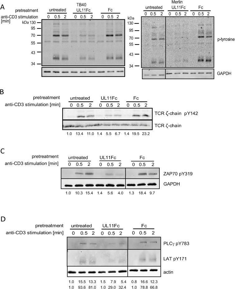
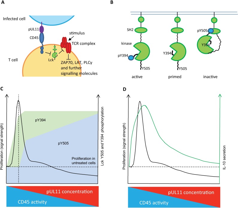
Human Cytomegalovirus (HCMV) is a widespread pathogen, infection with which can cause severe disease for immunocompromised individuals. The complex changes wrought on the host's immune system during both productive and latent HCMV infection are well known. Infected cells are masked and manipulated and uninfected immune cells are also affected; peripheral blood mononuclear cell (PBMC) proliferation is reduced and cytokine profiles altered. Levels increase of the anti-inflammatory cytokine IL-10, which may be important for the establishment of HCMV infections and is required for the development of high viral titres by murine cytomegalovirus. The mechanisms by which HCMV affects T cell IL-10 secretion are not understood. We show here that treatment of PBMC with purified pUL11 induces IL-10 producing T cells as a result of pUL11 binding to the CD45 phosphatase on T cells. IL-10 production induced by HCMV infection is also in part mediated by pUL11. Supernatants from pUL11 treated cells have anti-inflammatory effects on untreated PBMC. Considering the mechanism, CD45 can be a positive or negative regulator of TCR signalling, depending on its expression level, and we show that pUL11 also has concentration dependent activating or inhibitory effects on T cell proliferation and on the kinase function of the CD45 substrate Lck. pUL11 is therefore the first example of a viral protein that can target CD45 to induce T cells with anti-inflammatory properties. It is also the first HCMV protein shown to induce T cell IL-10 secretion. Understanding the mechanisms by which pUL11-induced changes in signal strength influence T cell development and function may provide the basis for the development of novel antiviral treatments and therapies against immune pathologies.


Conflict of interest statement

The authors have declared that no competing interests exist.

Figures

Fig 1
Fig 1. pUL11 treatment leads to the induction of IL-10 secretion.
A) PBMC were untreated (medium) or stimulated with anti-CD3 (OKT3) in the presence or absence of UL11Fc (TB40), UL11Fc (Merlin), UL6Fc or the Fc control protein at the indicated concentrations for 48h. B) PBMC were untreated or stimulated with anti-CD3 or treated with 50nM UL11Fc (TB40), UL11Fc (Merlin) or Fc without anti-CD3 (UL11Fc/Fc) or with anti-CD3, following preincubation with the indicated concentrations of anti-CD45 (AICD45.2) where shown, for 96h. C) Enriched primary T cells were untreated or stimulated with anti-CD3 and anti-CD28 in the presence or absence of UL11Fc (TB40) or Fc as indicated, for 48h. IL-10 secretion into the supernatant was measured by ELISA. Single representative experiments from two or three replicates are shown. Error bars show standard deviation of technical replicates.
Fig 2
Fig 2. HCMV infected epithelial cells induce IL-10 secretion from PBMC.
PBMC were incubated for 48h alone or with 50nM UL11Fc or Fc as a control, or with RPE cells that were uninfected (RPE) or infected with wild type HCMV (WT RPE) or ΔUL11 HCMV (ΔUL11 RPE) at the indicated ratios and in the absence or presence of anti-CD3 (OKT3). The presence of secreted IL-10 was measured by ELISA after 2 days. The experiment was performed three times, using PBMC from different donors. (A) One representative experiment is shown, with IL-10 concentration in pg/ml. Error bars show standard deviation of technical replicates. (B) Mean fold increases in IL-10 concentration over the value for uninfected RPE cells incubated with PBMC at a ratio of 1:5 in the presence of anti-CD3 are shown for the three experiments combined. Error bars show standard deviation. Statistical significance of differences between groups was determined by ANOVA. *p = 0.0005 for the difference in IL-10 expression between supernatants from cultures containing wild type HCMV and those containing ΔUL11 HCMV infected cells.
Fig 3
Fig 3. IL-10 secretion following pUL11 treatment accumulates over several days and involves the formation of new IL-10 producing T cells.
PBMC were left untreated (medium) or incubated with anti-CD3 (OKT3) or with anti-CD3 and UL11Fc (TB40), UL11Fc (Merlin) or Fc at the (A) indicated concentrations and times or for (B) 72h. (A) Supernatants were harvested and the concentration of IL-10 measured by ELISA after 3 days of incubation. One representative experiment out of two or three is shown. Error bars represent standard deviation of technical replicates. (B) Cells labelled extracellularly with anti-CD3 and anti-CD4, and intracellularly with anti-IL-10 were measured by flow cytometry. Percentages of live CD3+ CD4+ cells are shown (i) and the IL-10+ cells from this compartment (ii). One representative experiment out of five is shown.
Fig 4
Fig 4. Supernatant from pUL11 treated cells inhibits IFNγ production.
PBMC were incubated for 48h in the absence (w/o PHA) or presence of PHA alone (PHA) or together with conditioned medium from untreated PBMC (Con med) or from PBMC treated with anti-CD3 alone (Con anti-CD3) or with the addition of 50nM or 100nM of UL11Fc (Con UL11 50, Con UL11 100) or 50nM or 100nM of the Fc control (Con Fc 50, Con Fc 100). IFNγ concentration in the supernatants was measured by ELISA after 48h of incubation. (A) One representative experiment out of three is shown. Error bars show standard deviation of technical replicates. (B) Fold increase in IFNγ concentration in comparison to that from cells stimulated with PHA alone is shown for the three experiments combined (normalised to values for PHA stimulated cells). Background IFNγ signals present in the conditioned medium were subtracted. Error bars indicate standard deviation. Statistical significance of differences between groups was determined by ANOVA. *p = 0.0034 for the difference between IFNγ expression in supernatants from cultures incubated with conditioned medium from pUL11Fc treated cells and those incubated with conditioned medium from Fc treated cells.
Fig 5
Fig 5. pUL11Fc modulates T cell proliferation in a dose dependent manner.
PBMCs were either left unstimulated or stimulated with anti-CD3 (OKT3) together with the indicated concentrations of UL11Fc derived from HCMV strain (A) TB40 or (B) Merlin or with the Fc control protein. Single representative experiments from four (A) or two (B) replicates are shown; error bars show standard deviation of technical replicates.
Fig 6
Fig 6. Treatment with pUL11Fc affects phosphorylation of the regulatory tyrosines of Lck.
PBMC were incubated with anti-CD3 (OKT3) in the presence or absence of UL11Fc and Fc at the indicated concentrations for three days. Cells were then labelled extracellularly with anti-CD3 and anti-CD4 and intracellularly with anti-Lck pY505 or anti-Lck pY394. The experiment was repeated three times using cells from different donors. Mean fluorescence intensities are shown for Y505 (i) and Y394 (ii) phosphorylation of live CD3+CD4+ cells, fold increases in geometric mean fluorescent intensity normalised to anti-CD3 treated cells are indicated. A) depicts one representative experiment (values for UL11 treated cells). B) shows fold increases in phosphorylation over all three experiments for UL11Fc and Fc treated cells. Error bars indicate standard deviation. Statistical significance of differences between groups was determined by ANOVA. i)*p = 0.0014 for the difference in Y505 phosphorylation between pUL11Fc and Fc treated cells. ii)*p = 0.0043 for the difference in Y394 phosphorylation between pUL11Fc and Fc treated cells.
Fig 7
Fig 7. pUL11 reduces the activation of T cells.
Jurkat T cells were either untreated or preincubated with 800nM UL11Fc (TB40), UL11Fc (Merlin) and the Fc control protein (A) or UL11Fc (TB40) and the Fc control protein (B, C and D) and then stimulated with an anti-TCR antibody for the indicated times. Activation of TCR signalling was detected by immunoblotting using phospho-specific antibodies; (A) overall tyrosine phosphorylation, (B) TCR ζ-chain Y142 phosphorylation, (C) ZAP-70 Y319 phosphorylation and (D) PLCγ Y738 and LAT Y171 phosphorylation. Quantification of the phospho-specific signals was performed for panels B)-D) and is shown normalised to untreated, unstimulated cells, correlated with loading control intensity.
Fig 8
Fig 8. Model.
A) pUL11 on the surface of infected cells interacts with T cell CD45. The ability of CD45 to dephosphorylate regulatory tyrosine residues of Lck is reduced. This, in turn, affects the phosphorylation of ITAMs in the TCR complex and other downstream signalling molecules (either positively or negatively, depending on Lck activity). B) Phosphorylation of Lck regulatory tyrosines Y505 and Y394 determines its kinase activity. Y505 phosphorylation induces a closed, inactive state. Y394 phosphorylation is activating, by allowing substrates access to the kinase domain. C) PBMC proliferation in response to stimulation is dependent on the phosphorylation state of Lck Y394 (activating) and Y505 (inhibitory). Peak proliferation (vertical dashed line) is seen when pY394 is increased and diminishes as pY505 increases, inactivating Lck. At the highest levels of pY505, proliferation is inhibited (below the horizontal dashed line). As the concentration of pUL11 is increased, the ability of CD45 to dephosphorylate Lck decreases, shown by increased levels of pY394 and pY505. CD45 preferentially dephosphorylates pY505, hence higher amounts of pUL11 are required to affect Y505 phosphorylation. D) IL-10 secretion induced by pUL11 treatment appears to be influenced by changes in signal strength. T cell IL-10 production is influenced by changes to downstream signalling intermediates such as could be affected by pUL11 treatment.[31,47].

Similar articles

Cited by

References

    1. Cannon MJ, Schmid DS, Hyde TB. Review of cytomegalovirus seroprevalence and demographic characteristics associated with infection. Reviews in Medical Virology. 2010. pp. 202–213. doi: 10.1002/rmv.655 - DOI - PubMed
    1. Ludwig A, Hengel H. Epidemiological impact and disease burden of congenital cytomegalovirus infection in Europe. Euro Surveill. 2009;14: 26–32. - PubMed
    1. Ramanan P, Razonable RR. Cytomegalovirus infections in solid organ transplantation: a review. Infect Chemother. 2013;45: 260–71. doi: 10.3947/ic.2013.45.3.260 - DOI - PMC - PubMed
    1. Hebart H, Einsele H. Clinical aspects of CMV infection after stem cell transplantation. Human Immunology. 2004. pp. 432–436. doi: 10.1016/j.humimm.2004.02.022 - DOI - PubMed
    1. Randolph-Habecker J, Iwata M, Torok-Storb B. Cytomegalovirus mediated myelosuppression. J Clin Virol. 2002;25 Suppl 2: S51–6. - PubMed

MeSH terms