. 2017 Jun 19;13(6):e1006456.
doi: 10.1371/journal.ppat.1006456. eCollection 2017 Jun.

The ApaH-like phosphatase TbALPH1 is the major mRNA decapping enzyme of trypanosomes

Affiliations

The ApaH-like phosphatase TbALPH1 is the major mRNA decapping enzyme of trypanosomes

Susanne Kramer. PLoS Pathog. .

Abstract

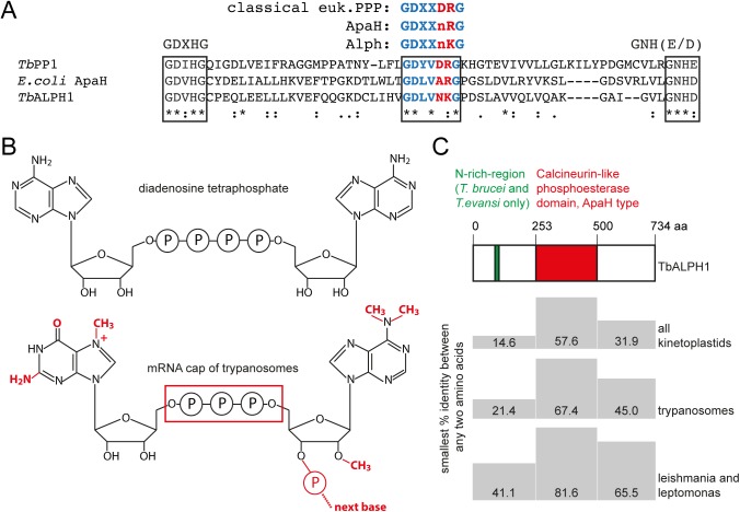
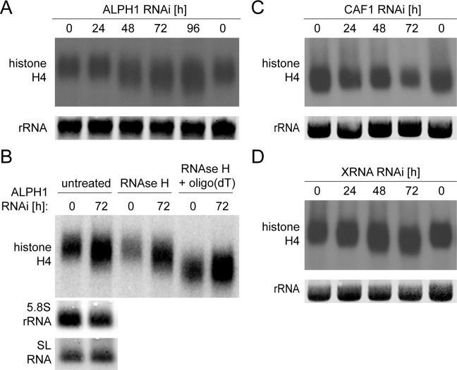
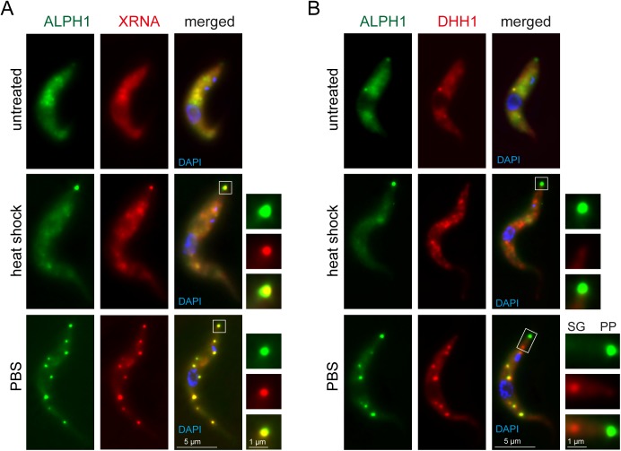
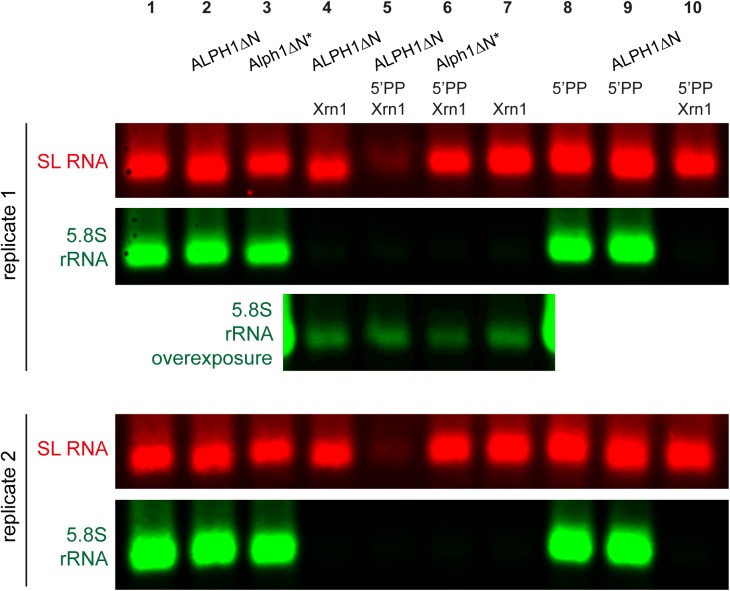
5'-3' decay is the major mRNA decay pathway in many eukaryotes, including trypanosomes. After deadenylation, mRNAs are decapped by the nudix hydrolase DCP2 of the decapping complex and finally degraded by the 5'-3' exoribonuclease. Uniquely, trypanosomes lack homologues to all subunits of the decapping complex, while deadenylation and 5'-3' degradation are conserved. Here, I show that the parasites use an ApaH-like phosphatase (ALPH1) as their major mRNA decapping enzyme. The protein was recently identified as a novel trypanosome stress granule protein and as involved in mRNA binding. A fraction of ALPH1 co-localises exclusively with the trypanosome 5'-3' exoribonuclease XRNA to a special granule at the posterior pole of the cell, indicating a connection between the two enzymes. RNAi depletion of ALPH1 is lethal and causes a massive increase in total mRNAs that are deadenylated, but have not yet started 5'-3' decay. These data suggest that ALPH1 acts downstream of deadenylation and upstream of mRNA degradation, consistent with a function in mRNA decapping. In vitro experiments show that recombinant, N-terminally truncated ALHP1 protein, but not a catalytically inactive mutant, sensitises the capped trypanosome spliced leader RNA to yeast Xrn1, but only if an RNA 5' polyphosphatase is included. This indicates that the decapping mechanism of ALPH1 differs from the decapping mechanism of Dcp2 by leaving more than one phosphate group at the mRNA's 5' end. This is the first reported function of a eukaryotic ApaH-like phosphatase, a bacterial-derived class of enzymes present in all phylogenetic super-groups of the eukaryotic kingdom. The substrates of eukaryotic ApaH-like phosphatases are unknown. However, the substrate of the related bacterial enzyme ApaH, diadenosine tetraphosphate, is highly reminiscent of a eukaryotic mRNA cap.


Conflict of interest statement

The authors have declared that no competing interests exist.

Figures

Fig 1
Fig 1. T. brucei ALPH1.
(A) Alignment of the signature motifs of a classical eukaryotic PPP (TbPP1, Tb927.4.3560) and the atypical PPPs ApaH of E. coli and ALPH1 of T. brucei. The three catalytic signature motifs of PPPs are framed. The two changes in the GDXXDRG motif that are characteristic for ApaH and Alph are highlighted in red. In Alph’s and ApaH, the second aspartate is replaced by a neutral amino acid (n). In most Alphs, but not in ApaH or other PPPs, arginine, a coordinator of phosphate [72], is replaced by lysine [41]. (B) Structure of diadenosine tetraphosphate (top) and of the mRNA cap of trypanosomes (bottom). The differences are marked in red. (C) Domain structure of ALPH1 and conservation between the different kinetoplastida orthologues. All available kinetoplastid orthologues to TbALPH1, (13 sequences of trypanosomes, 1 of Leptomonas and 6 of Leishmania strains) were aligned by ClustalW using default parameters. The minimal % identity between any two sequences is indicated for the three different ALPH domains. The sequences of trypanosomes and Leishmania/Leptomonas were also analysed separately.
Fig 2
Fig 2. RNAi depletion of ALPH1 causes growth arrest and increase in mRNA levels.
RNAi depletion of TbALPH1 was induced by tetracycline (TET). Three independent clonal cell lines were analysed over a time-course of RNAi induction. (A) Reduction in the number of ALPH1 mRNA molecules. ALPH1 mRNA and DBP1 mRNA (control) were visualized by dual colour single molecule mRNA FISH (Affymetrix), using green (ALPH1) and red (DBP1) fluorescent Affymetrix probe sets. The number of mRNA molecules per cell was quantified after 0, 24 and 48 hours of ALPH1 RNAi induction. Data are presented as box plot (waist is median; box is IQR; whiskers are ±1.5 IQR; only the smallest and largest outliers are shown; n = 100 for each time-point); the number of mRNAs from the individual cells is also presented as green (ALPH1) or red (DBP1) dots. One representative cell for each time-point is shown. The data are from one RNAi clone; data of a second clone are shown in S12 Fig. (B) Growth arrest. Growth was measured over a time-course of ALPH1 RNAi induction (±TET). Averages of the three clonal cell lines are shown; error bars indicate standard deviations between the three cell lines. (C-E) Increase in mRNAs. Total RNA was isolated over a time-course of ALPH1 RNAi and as a control from bloodstream form trypanosomes (BSF) and analysed by northern blots. The blots were probed for total mRNA with an oligo antisense to the miniexon of the spliced leader RNA (C), for PGKC (D) and for GPI-PLC (E). mRNA abundances were quantified by Odyssey (total mRNA) or phosphorimager (PGKC, GPI-PLC). rRNA was used as a loading control and all samples were calibrated to the amount of BSF RNA (= 1). Average values of the three clones are shown, standard deviations are presented as error bars. For each probe, one representative northern blot is shown. Note that the three red bands in C) are not rRNA bands, but are caused by a squeezing of the mRNAs due to the very abundant rRNA.
Fig 3
Fig 3. mRNAs that accumulate after ALPH1 RNAi are deadenylated.
Northern blots were probed for histone H4, a very small mRNA that allows to visualise changes in poly(A) tail length by band shifts. (A) RNA was isolated over a time-course of ALPH1 RNAi depletion. Representative data from one of three RNAi clones are shown. (B) RNA isolated after 0 or 72 hours of ALPH1 RNAi depletion was treated with RNAse H in the absence or presence of oligo dT. Samples not treated with RNAse H served as control. The blot was re-probed for two small RNAs that have no poly(A) tail (5.8S rRNA and SL RNA) to demonstrate that the band-shift is specific to polyadenylated RNAs. (C) RNA was isolated over a time-course of CAF1 RNAi depletion. Representative data from one of three RNAi clones are shown. (D) RNA was isolated over a time-course of XRNA RNAi depletion. Representative data from one of two RNAi clones are shown.
Fig 4
Fig 4. RNAi depletion of ALPH1 causes an increase in intact mRNA molecules, but no increase in 5’-3’ decay intermediates.
(A) Experimental design: The endogenous very long and short-lived mRNA Tb427.01.1740 was used as a reporter for mRNA metabolism [55]. Simultaneous probing of the extreme 5’ and 3’ ends with red and green fluorescent single mRNA FISH probes results in yellow, green or red fluorescent mRNA molecules, corresponding to intact mRNAs, mRNAs in 5’-3’ decay and mRNAs in transcription or 3’-5’ decay, respectively. (B) The numbers of yellow, green and red fluorescent spots were quantified from cells after induction of ALPH1 RNAi (0, 24 or 48 hours TET) from at least 130 cells per time-point, for three RNAi clones. Average data are shown; error bars represent the standard deviations between the different RNAi clones. The numbers of the differently coloured spots per total cell are shown (left), but also the numbers of spots in the nucleus (middle) or in the cytoplasm (right). If the centre of a spot was overlapping with the DAPI stained nucleus on a Z-stack projection image, the spot was defined as nuclear, otherwise as cytoplasmic. (C) Representative Z-stack projection images of untreated cells (no TET) and cells after 48 hours of ALPH1 RNAi (48 h TET).
Fig 5
Fig 5. Only XRNA fully co-localises with ALPH1.
(A) Cells co-expressing XRNA-mChFP and ALPH1-eYFP from endogenous loci were treated with heat shock or starvation (PBS). The posterior pole granule is shown enlarged. (B) Cells co-expressing mChFP-DHH1 and ALPH1-eYFP from endogenous loci were treated with heat shock or starvation (PBS). The posterior pole granule (PP) is shown enlarged; for the PBS-treated cells the enlargement also includes the most posterior stress granule (SG) to show the absence of ALPH1.
Fig 6
Fig 6. ALPH1ΔN has in vitro decapping activity, in the presence of an RNA 5’ polyphosphatase.
Total trypanosome mRNA was treated with recombinant ALPH1ΔN or Alph1ΔN* as indicated, purified, treated with RNA 5’ polyphosphatase as indicated, purified, and finally treated with yeast Xrn1 as indicated. The RNA samples were analysed by northern blot probed for the SL RNA (red) and the 5.8S rRNA (green). For lane 4–7 of replicate 1, an overexposed blot of the 5.8S rRNA is shown to demonstrate equal loading. Data of two representative replicates are shown, out of a total of four replicates.

References

    1. Decker CJ, Parker R. P-bodies and stress granules: possible roles in the control of translation and mRNA degradation. Cold Spring Harb Perspect Biol. 2012. September;4(9):a012286 doi: 10.1101/cshperspect.a012286 - DOI - PMC - PubMed
    1. Courchaine EM, Lu A, Neugebauer KM. Droplet organelles? EMBO J. 2016. June 29;:e201593517. - PMC - PubMed
    1. Kramer S. RNA in development: how ribonucleoprotein granules regulate the life cycles of pathogenic protozoa. WIREs RNA. 2014. March;5(2):263–84. doi: 10.1002/wrna.1207 - DOI - PubMed
    1. Lykke-Andersen J. Identification of a Human Decapping Complex Associated with hUpf Proteins in Nonsense-Mediated Decay. Mol Cell Biol. 2002. December 1;22(23):8114–21. doi: 10.1128/MCB.22.23.8114-8121.2002 - DOI - PMC - PubMed
    1. Steiger M, Carr-Schmid A, Schwartz DC, Kiledjian M, Parker R. Analysis of recombinant yeast decapping enzyme. RNA. 2003. February;9(2):231–8. doi: 10.1261/rna.2151403 - DOI - PMC - PubMed

MeSH terms