Review
. 2017 Jun 18;18(6):1296.
doi: 10.3390/ijms18061296.

Cutaneous Manifestations of Human and Murine Leishmaniasis

Affiliations
Review

Cutaneous Manifestations of Human and Murine Leishmaniasis

Breanna M Scorza et al. Int J Mol Sci. .

Abstract

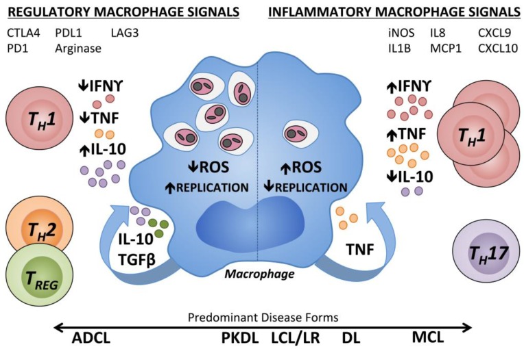
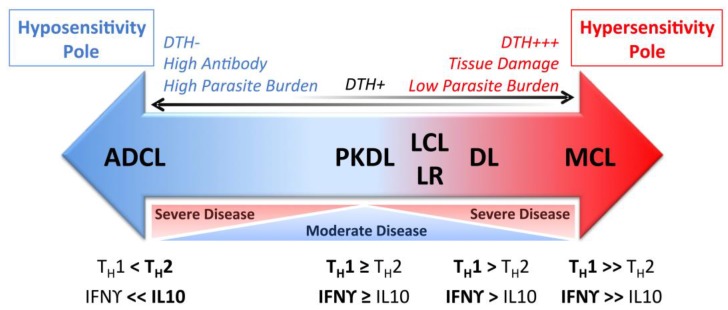
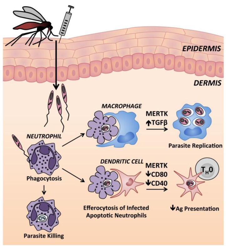
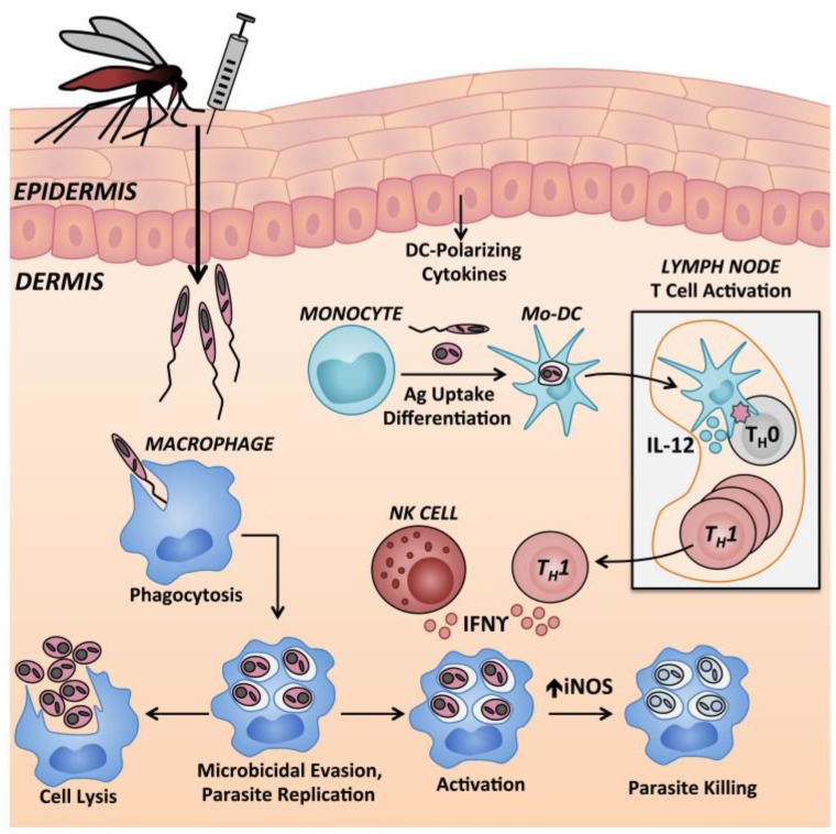
The leishmaniases are diseases caused by pathogenic protozoan parasites of the genus Leishmania. Infections are initiated when a sand fly vector inoculates Leishmania parasites into the skin of a mammalian host. Leishmania causes a spectrum of inflammatory cutaneous disease manifestations. The type of cutaneous pathology is determined in part by the infecting Leishmania species, but also by a combination of inflammatory and anti-inflammatory host immune response factors resulting in different clinical outcomes. This review discusses the distinct cutaneous syndromes described in humans, and current knowledge of the inflammatory responses associated with divergent cutaneous pathologic responses to different Leishmania species. The contribution of key hematopoietic cells in experimental cutaneous leishmaniasis in mouse models are also reviewed and compared with those observed during human infection. We hypothesize that local skin events influence the ensuing adaptive immune response to Leishmania spp. infections, and that the balance between inflammatory and regulatory factors induced by infection are critical for determining cutaneous pathology and outcome of infection.

Keywords: DTH-delayed type hypersensitivity skin test; LST-Leishmania skin test; Leishmania; cutaneous leishmaniasis; diffuse leishmaniasis; disseminated leishmaniasis; kala-azar; mucosal leishmaniasis; post-kala-azar dermal leishmaniasis; skin immunology.


Conflict of interest statement

The authors declare no conflict of interest.

Figures

Figure 1
Figure 1
Immune regulation of human macrophages contributing to different pathologic states during Leishmania infection. Human macrophages parasitized by Leishmania spp. are subject to regulation by cytokines present within the skin at the infection site. Inflammatory Type 1 cytokines, IFNγ and TNF, can synergistically induce ROS production by macrophages, thereby inhibiting Leishmania replication within the phagolysosome. TH17 cells are increased in inflammatory MCL lesions. Chemoattractant transcripts (IL8, MCP1, CXCL9, and CXCL10) and transcripts associated with classical (type 1) macrophage activation (iNOS, IL1β) have been measured in inflammatory CL lesions. In a regulatory environment (left), there are low levels of type 1 cytokines with lack of microbicidal effectors. T cell or macrophage-derived cytokines including type 2 cytokines, IL-10 and TGFβ antagonize the effects of IFNγ and TNF, and enhanced polyamines can result in parasite proliferation. TREG cells can suppress the effects of IFNγ. Inhibitory receptors on CD4+ or CD8+ T cells (CTLA4, PD1, and LAG3) and their counter-ligands (CD80, CD86, and PDL1) are associated with T cell exhaustion. Arginase activity is associated with M2-type non-classical macrophage activation. IFNγ: Interferon γ; TNF: Tumor necrosis factor; IL10: Interleukin 10; TGFβ: Transforming growth factor β; ROS: Reactive oxygen species; ADCL: Anergic diffuse cutaneous leishmaniasis; PKDL: Post Kala-Azar dermal leishmaniasis; LCL: Localized cutaneous leishmaniasis; LR: Leishmania recidivans; DL: Disseminated leishmaniasis; MCL: Mucocutaneous leishmaniasis; DTH: Delayed type 1 hypersensitivity; TREG: T regulatory cell; TH: T helper cell; MCP: Monocyte chemoattractant protein; CXCL: Chemokine (C-X-C motif) ligand; CTLA4: Cytotoxic T-lymphocyte associated protein 4; PD1: Programmed cell death protein 1; LAG3: Lymphocyte activation gene 3; PDL1: Programmed death ligand 1.
Figure 2
Figure 2
Schematic representation of the spectrum of human cutaneous leishmaniasis manifestations. The localized disease LCL results in the presence of an IFNγ-dominated response, detected by DTH response and T cells. In MCL, T cell responsiveness to Leishmania antigen further increases towards the hypersensitivity pole, resulting in tissue damage but low parasite burden. The opposite polar responses with low or absent T cell responsiveness, high parasite burden and antibody titers culminate in ADCL. ADCL: Anergic diffuse cutaneous leishmaniasis; PKDL: Post Kala-Azar dermal leishmaniasis; LCL: Localized cutaneous leishmaniasis; LR: Leishmania recidivans; DL: Disseminated leishmaniasis; MCL: Mucocutaneous leishmaniasis; DTH: Delayed type 1 hypersensitivity; TH: T helper cell; TH1: TH1-type immune response; TH2: TH2-type immune response; IFNγ: Interferon gamma; IL-10: Interleukin 10.
Figure 3
Figure 3
Neutrophils at the cutaneous site of Leishmania spp. inoculation in mice. Neutrophils are rapidly recruited to the site of Leishmania infection, initiated by either sand fly or needle inoculation, where they phagocytose parasites. Although some Leishmania are killed by neutrophils, some survive within neutrophils until they undergo apoptosis. Uptake of apoptotic infected neutrophils by macrophages leads to MERTK signaling and increased TGFβ release, promoting an anti-inflammatory macrophage state and intracellular parasite replication. DCs that take up apoptotic infected neutrophils show a decreased capacity to present antigen to naïve CD4+ T cells. MERTK: Mer tyrosine kinase; TGFβ: Transforming growth factor β, TH0: naïve CD4+ T cell.
Figure 4
Figure 4
Monocytic cells in control of Leishmania replication in murine skin. Epidermal cytokines can modulate this process. Monocyte derived dendritic cells (Mo-DCs) transport antigen to draining lymph nodes for presentation to naïve CD4+ T cells and resultant TH1 cells traffic back to the site of infection where they secrete IFNγ. Recruited inflammatory monocytes are the primary host cell allowing intracellular survival and replication of Leishmania spp. Monocytes recruited to the site of intradermal Leishmania inoculation can differentiate locally into dendritic cells or macrophages. Once phagocytosed by macrophages, Leishmania use a variety of virulence mechanisms to suppress the host cell microbicidal response and replicate intracellularly. Infected macrophages can either facilitate intracellular survival and/or replication, or if exposed to activating signals such as IFNγ from TH1-type cells or NK cells, can upregulate iNOS. The combination of reactive oxygen and reactive nitrogen species can contribute to killing of intracellular parasites. Mo-DC: Monocyte derived dendritic cell; NK cell: Natural killer cell; IFNγ: Interferon γ; IL-12: Interleukin-12; iNOS: Inducible nitric oxide synthase; TH1: T helper 1 cell, TH0: Naïve CD4+ T cell.

References

    1. WHO Weekly Epidemiological Record. [(accessed on 31 August 2016)]; Available online: http://www.who.int/wer/2016/wer9122.pdf?ua=1.
    1. Grinnage-Pulley T., Scott B., Petersen C.A. A mother’s gift: Congenital transmission of trypanosoma and leishmania species. PLoS Pathog. 2016;12:e1005302. doi: 10.1371/journal.ppat.1005302. - DOI - PMC - PubMed
    1. Pearson R.D., Sousa A.Q. Clinical spectrum of leishmaniasis. Clin. Infect. Dis. 1996;22:1–13. doi: 10.1093/clinids/22.1.1. - DOI - PubMed
    1. Malaviya P., Picado A., Singh S.P., Hasker E., Singh R.P., Boelaert M., Sundar S. Visceral leishmaniasis in Muzaffarpur district, Bihar, India from 1990 to 2008. PLoS ONE. 2011;6:e14751. - PMC - PubMed
    1. Al-Salem W.S., Pigott D.M., Subramaniam K., Haines L.R., Kelly-Hope L., Molyneux D.H., Hay S.I., Acosta-Serrano A. Cutaneous leishmaniasis and conflict in Syria. Emerg. Infect. Dis. 2016;22:931–933. doi: 10.3201/eid2205.160042. - DOI - PMC - PubMed

LinkOut - more resources