. 2017 Jun 19;9(1):57.
doi: 10.1186/s13073-017-0447-8.

Linked read sequencing resolves complex genomic rearrangements in gastric cancer metastases

Affiliations

Linked read sequencing resolves complex genomic rearrangements in gastric cancer metastases

Stephanie U Greer et al. Genome Med. .

Abstract

Background: Genome rearrangements are critical oncogenic driver events in many malignancies. However, the identification and resolution of the structure of cancer genomic rearrangements remain challenging even with whole genome sequencing.

Methods: To identify oncogenic genomic rearrangements and resolve their structure, we analyzed linked read sequencing. This approach relies on a microfluidic droplet technology to produce libraries derived from single, high molecular weight DNA molecules, 50 kb in size or greater. After sequencing, the barcoded sequence reads provide long range genomic information, identify individual high molecular weight DNA molecules, determine the haplotype context of genetic variants that occur across contiguous megabase-length segments of the genome and delineate the structure of complex rearrangements. We applied linked read sequencing of whole genomes to the analysis of a set of synchronous metastatic diffuse gastric cancers that occurred in the same individual.

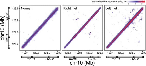
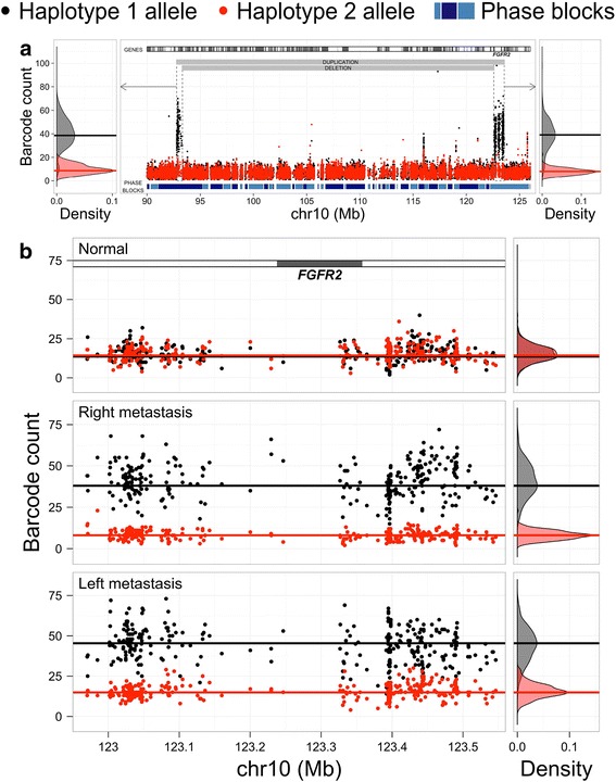
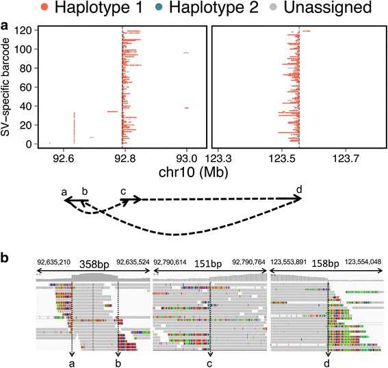
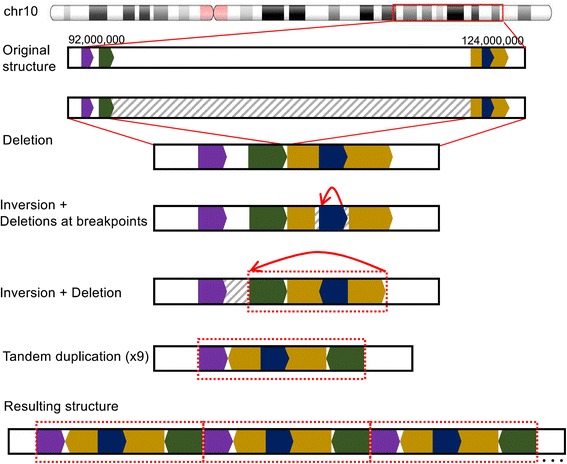
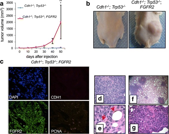
Results: When comparing metastatic sites, our analysis implicated a complex somatic rearrangement that was present in the metastatic tumor. The oncogenic event associated with the identified complex rearrangement resulted in an amplification of the known cancer driver gene FGFR2. With further investigation using these linked read data, the FGFR2 copy number alteration was determined to be a deletion-inversion motif that underwent tandem duplication, with unique breakpoints in each metastasis. Using a three-dimensional organoid tissue model, we functionally validated the metastatic potential of an FGFR2 amplification in gastric cancer.

Conclusions: Our study demonstrates that linked read sequencing is useful in characterizing oncogenic rearrangements in cancer metastasis.

Keywords: Barcode linked sequence reads; Cancer drivers; Cancer rearrangements; High molecular weight DNA; Whole genome analysis.


Figures

Fig. 1
Fig. 1
Barcode overlap plots of the genomic region surrounding the proto-oncogene FGFR2. The level of barcode sharing between 10-kb windows in a 1.4-Mb genomic region including FGFR2 was determined for the normal sample and the right and left metastatic samples. The highest level of overlap (red) is expected along the diagonal, while off-diagonal signals (red or blue) indicate the presence of structural variants
Fig. 2
Fig. 2
Allele-specific barcode counts. a For the right metastasis, the number of barcodes associated with each allele of all phased heterozygous variants is shown for a 36-Mb genomic region including FGFR2. The allelic barcode counts are colored in black and red to denote belonging to haplotype 1 or haplotype 2 within each phase block. The locations of the duplication and deletion events, as identified by Long Ranger, are indicated. The barcode count densities are plotted for each amplified region before and after the deletion event (regions denoted by dashed rectangles). b Allele-specific barcode counts for each phased allele in the tumor-amplified region of FGFR2, using the normal sample to define allelic assignment to haplotype 1 (black) or haplotype 2 (red). The same haplotype (haplotype 1; black) is amplified in both metastases
Fig. 3
Fig. 3
Complex breakpoint resolution using molecular barcode mapping. a The SV-specific molecules for breakpoint 1 and breakpoint 2 of the duplication SV in the right metastasis are plotted according to the mapping location of molecular barcoded reads. Each row of the plot represents one SV-specific molecule, depicting how each SV-specific molecule spans the SV breakpoint. Molecular breakpoints are denoted with a, b, c, and d, and the arrow structure indicates breakpoint connection and directionality. b IGV plots of the molecular breakpoints display soft-clip evidence of the breakpoints
Fig. 4
Fig. 4
Putative structural rearrangement of the FGFR2 genomic region in the right metastasis. Barcode and read-based evidence indicate the likely occurrence of events was a 30-Mb deletion event with a nearby inversion event, and an inversion event with a deletion at the boundary; the resulting rearrangement then underwent an approximately ninefold tandem duplication. Barcode analysis indicates that all of these events are in cis with one another and thus occurred on only one copy of chromosome 10
Fig. 5
Fig. 5
Gastric organoid tumor model. Gastric organoids with the indicated genotypes are shown. a Tumor volumes were measured over time post-injection. Gastric organoids were dissociated and subcutaneously injected into the flanks of NOG mice. Cdh1 -/-;Trp53 -/- is shown in blue, and Cdh1 -/-;Trp53 -/-;FGFR2 is shown in red. Error bars represent SEM, and asterisks indicate p < 0.04. b Images indicate tumor growth at 50 days post-injection. c Overexpression of FGFR2 was confirmed in the tumor derived from Cdh1 -/-;Trp53 -/-;FGFR2 organoids. de Histological analysis of the Cdh1-/-;Trp53-/-;FGFR2 tumors confirms the presence of poorly differentiated adenocarcinoma with signet ring as indicated by arrows. f, g After flank injections with dissociated organoids, histological analysis of murine lungs after 50 days revealed metastatic gastric adenocarcinoma with signet ring features at low (f) and high (g) magnification

References

    1. Sudmant PH, Rausch T, Gardner EJ, Handsaker RE, Abyzov A, Huddleston J, Zhang Y, Ye K, Jun G, Hsi-Yang Fritz M, et al. An integrated map of structural variation in 2,504 human genomes. Nature. 2015;526:75–81. doi: 10.1038/nature15394. - DOI - PMC - PubMed
    1. Weischenfeldt J, Symmons O, Spitz F, Korbel JO. Phenotypic impact of genomic structural variation: insights from and for human disease. Nat Rev Genet. 2013;14:125–38. doi: 10.1038/nrg3373. - DOI - PubMed
    1. Yang L, Luquette LJ, Gehlenborg N, Xi R, Haseley PS, Hsieh CH, Zhang C, Ren X, Protopopov A, Chin L, et al. Diverse mechanisms of somatic structural variations in human cancer genomes. Cell. 2013;153:919–29. doi: 10.1016/j.cell.2013.04.010. - DOI - PMC - PubMed
    1. Rowley JD. Letter: A new consistent chromosomal abnormality in chronic myelogenous leukaemia identified by quinacrine fluorescence and Giemsa staining. Nature. 1973;243:290–3. doi: 10.1038/243290a0. - DOI - PubMed
    1. Nowell PC, Hungerford DA. Chromosome studies on normal and leukemic human leukocytes. J Natl Cancer Inst. 1960;25:85–109. - PubMed

Publication types