Comparative Study
. 2017 Aug 25;292(34):14240-14249.
doi: 10.1074/jbc.M117.798405. Epub 2017 Jun 19.

The glutamate/cystine antiporter SLC7A11/xCT enhances cancer cell dependency on glucose by exporting glutamate

Affiliations
Comparative Study

The glutamate/cystine antiporter SLC7A11/xCT enhances cancer cell dependency on glucose by exporting glutamate

Pranavi Koppula et al. J Biol Chem. .

Abstract

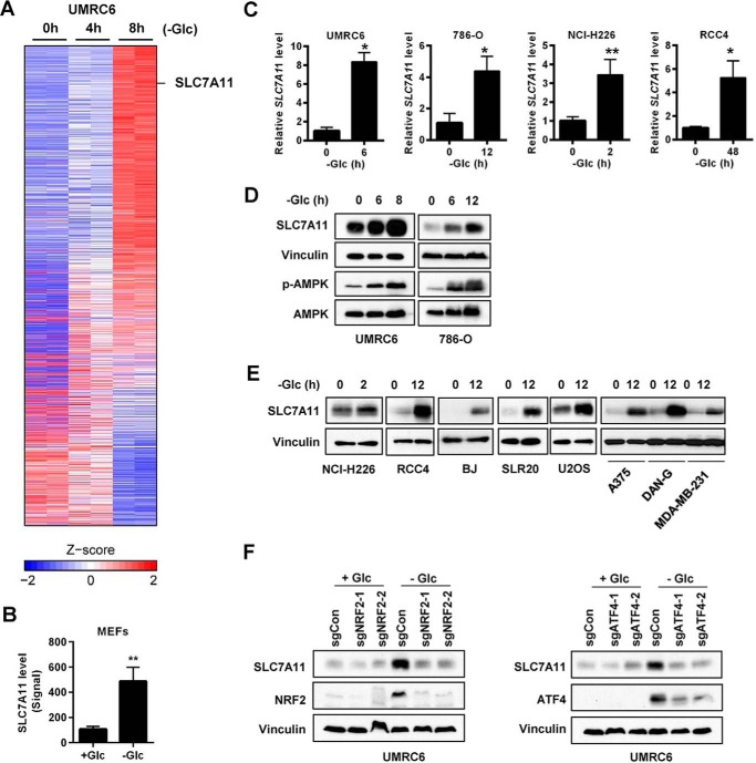
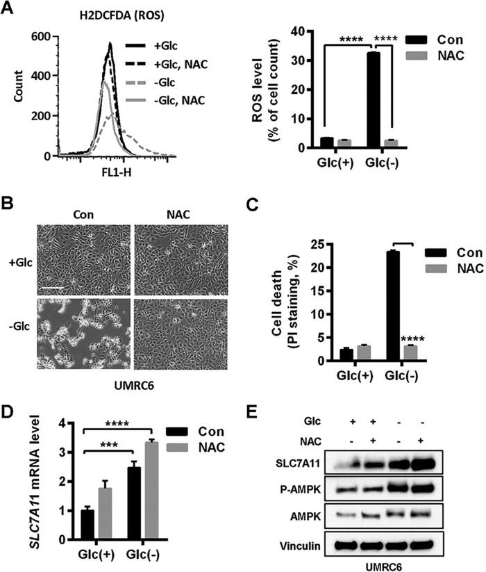
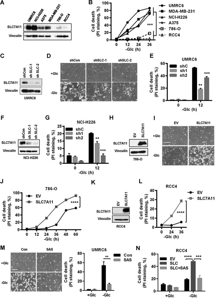
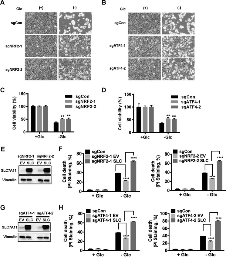
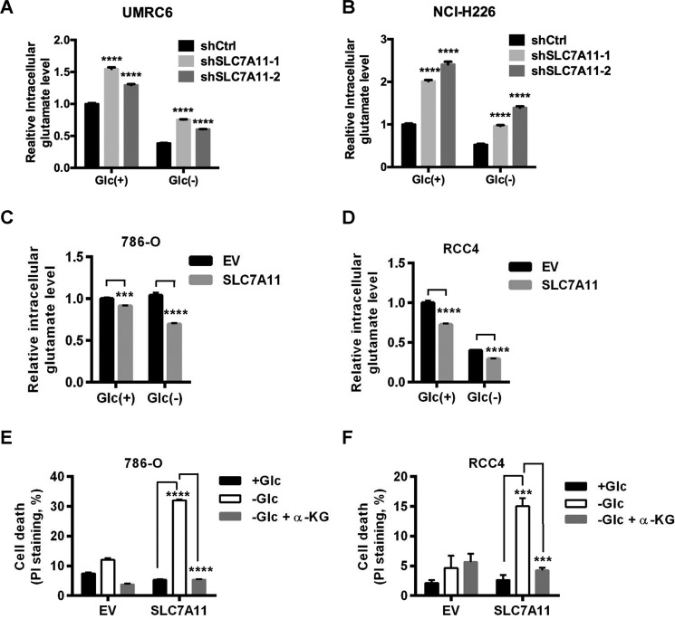
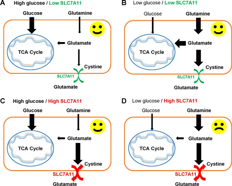
Cancer cells with specific genetic alterations may be highly dependent on certain nutrients for survival, which can inform therapeutic strategies to target these cancer-specific metabolic vulnerabilities. The glutamate/cystine antiporter solute carrier family 7 member 11 (SLC7A11, also called xCT) is overexpressed in several cancers. Contrasting the established pro-survival roles of SLC7A11 under other stress conditions, here we report the unexpected finding that SLC7A11 overexpression enhances cancer cell dependence on glucose and renders cancer cells more sensitive to glucose starvation-induced cell death and, conversely, that SLC7A11 deficiency by either knockdown or pharmacological inhibition promotes cancer cell survival upon glucose starvation. We further show that glucose starvation induces SLC7A11 expression through ATF4 and NRF2 transcription factors and, correspondingly, that ATF4 or NRF2 deficiency also renders cancer cells more resistant to glucose starvation. Finally, we show that SLC7A11 overexpression decreases whereas SLC7A11 deficiency increases intracellular glutamate levels because of SLC7A11-mediated glutamate export and that supplementation of α-ketoglutarate, a key downstream metabolite of glutamate, fully restores survival in SLC7A11-overexpressing cells under glucose starvation. Together, our results support the notion that both glucose and glutamate have important roles in maintaining cancer cell survival and uncover a previously unappreciated role of SLC7A11 to promote cancer cell dependence on glucose. Our study therefore informs therapeutic strategies to target the metabolic vulnerability in tumors with high SLC7A11 expression.

Keywords: SLC7A11; cell death; glucose; glutamate; reactive oxygen species (ROS); tumor metabolism; xCT.


Conflict of interest statement

The authors declare that they have no conflicts of interest with the contents of this article

Figures

Figure 1.
Figure 1.
Glucose starvation induces SLC7A11 through ATF4 and NRF2. A, heatmap representation of genes regulated upon glucose (Glc) starvation in UMRC6 cells. B, normalized SLC7A11 level from a mouse microarray using mouse embryonic fibroblasts (MEFs) treated with and without glucose for 24 h. C–E, glucose starvation–induced SLC7A11 expression in different cells was analyzed by RT-PCR (C) and Western blot analysis (D and E). F, the effects of NRF2 and ATF4 deletion on SLC7A11 induction upon glucose starvation in UMRC6 cells were examined by Western blotting. *, p < 0.05; **, p < 0.01.
Figure 2.
Figure 2.
Glucose starvation induces SLC7A11 independent of ROS. A, cellular ROS levels in UMRC6 cells after glucose (Glc) withdrawal for 6 h were determined by H2DCFDA flow cytometer analysis. To prevent ROS generation, 2 mm NAC was added to the glucose-free medium. Right panel, data represent mean ± S.D. (n = 3). Con, control. B and C, UMRC6 cells cultured in glucose-free medium for 6 h were monitored under inverted microscopy (B), and cell death was analyzed by PI staining (C). Scale bar = 100 μm. D and E, SLC7A11 mRNA and protein levels in UMRC6 cells upon glucose starvation and NAC treatment were analyzed by RT-PCR (D) and Western blotting (E). ***, p < 0.001; ****, p < 0.0001.
Figure 3.
Figure 3.
SLC7A11 sensitizes cancer cells to glucose starvation–induced cell death. A, SLC7A11 protein levels in different cancer cell lines were determined by Western blotting. B, cell death upon glucose (Glc) starvation for 0–36 h in different cancer cell lines was analyzed by PI staining. C, SLC7A11 protein levels in SLC7A11 knockdown UMRC6 cells. D and E, cell death in SLC7A11 knockdown UMRC6 cells upon glucose withdrawal was monitored (D) and analyzed by PI staining (E). Scale bar = 100 μm. F, SLC7A11 protein levels in SLC7A11 knockdown NCI-H226 cells. G, cell death upon glucose withdrawal was analyzed by PI staining for SLC7A11 knockdown NCI-H226 cells. H, SLC7A11 protein levels in SLC7A11-overexpressing 786-O cells. EV, empty vector. I and J, cell death in SLC7A11-overexpressing 786-O cells upon glucose withdrawal was monitored (I) and analyzed by PI staining (J). Scale bar = 100 μm. K, SLC7A11 protein levels in SLC7A11-overexpressing RCC4 cells. L, cell death in SLC7A11-overexpressing RCC4 cells upon glucose withdrawal was analyzed by PI staining. M and N, cell death in UMRC6 cells (M) or SLC7A11-overexpressing RCC4 cells (N) upon glucose withdrawal and treatment with 1 mm sulfasalazine (SAS) was monitored and analyzed by PI staining. **, p < 0.01; ***, p < 0.001; ****, p < 0.0001.
Figure 4.
Figure 4.
ATF4 and NRF2 promote glucose starvation–induced cell death. A–D, NRF2- and ATF4-deficient cells were cultured in glucose (Glc)-free medium for 6 h. Shown are representative images (A and B) and quantification of cell viability (C and D) of the indicated cell lines that were cultured in glucose-free medium for 6 h. Scale bar = 100 μm. E and G, SLC7A11 protein levels in SLC7A11-overexpressing NRF2-deficient (E) or ATF4-deficient cells (G). EV, empty vector. F and H, cell death in UMRC6 cells with the indicated genotypes after 5-h glucose starvation was analyzed by PI staining. **, p < 0.01; ****, p < 0.0001.
Figure 5.
Figure 5.
SLC7A11 regulation of glutamate efflux underlies SLC7A11-mediated increased sensitivity to glucose starvation. A and B, intracellular glutamate levels were determined in SLC7A11 knockdown UMRC6 cells after 3 h of glucose (Glc) withdrawal (A) or NCI-H226 cells after 1 h of glucose withdrawal (B). All values were normalized to control shRNA (shCon) cells in complete medium with 25 mm glucose. C and D, intracellular glutamate levels were determined in SLC7A11-overexpressing 786-O cells after 8 h of glucose withdrawal (C) or RCC4 cells after 14 h of glucose withdrawal (D). All values were normalized to empty vector (EV) cells in complete medium with 25 mm glucose. E and F, SLC7A11-overexpressing 786-O cells (E) or RCC4 cells (F) were cultured in complete medium or glucose-free medium with and without 5 mm dimethyl-αKG for 24 h. Cell death was measured by PI staining. ***, p < 0.001; ****, p < 0.0001.
Figure 6.
Figure 6.
Model for the role of SLC7A11 in glucose dependence. See “Discussion” for a detailed description.

References

    1. Vander Heiden M. G., Cantley L. C., and Thompson C. B. (2009) Understanding the Warburg effect: the metabolic requirements of cell proliferation. Science 324, 1029–1033 - PMC - PubMed
    1. Vander Heiden M. G., and DeBerardinis R. J. (2017) Understanding the intersections between metabolism and cancer biology. Cell 168, 657–669 - PMC - PubMed
    1. DeBerardinis R. J., and Chandel N. S. (2016) Fundamentals of cancer metabolism. Sci. Adv. 2, e1600200. - PMC - PubMed
    1. Ward P. S., and Thompson C. B. (2012) Metabolic reprogramming: a cancer hallmark even Warburg did not anticipate. Cancer Cell 21, 297–308 - PMC - PubMed
    1. Hensley C. T., Wasti A. T., and DeBerardinis R. J. (2013) Glutamine and cancer: cell biology, physiology, and clinical opportunities. J. Clin. Invest. 123, 3678–3684 - PMC - PubMed

Publication types

MeSH terms