. 2017 Aug 18;85(9):e00176-17.
doi: 10.1128/IAI.00176-17. Print 2017 Sep.

Pseudomonas Quinolone Signal Induces Oxidative Stress and Inhibits Heme Oxygenase-1 Expression in Lung Epithelial Cells

Affiliations

Pseudomonas Quinolone Signal Induces Oxidative Stress and Inhibits Heme Oxygenase-1 Expression in Lung Epithelial Cells

Maher Y Abdalla et al. Infect Immun. .

Abstract

Pseudomonasaeruginosa causes lung infections in patients with cystic fibrosis (CF). The Pseudomonas quinolone signal (PQS) compound is a secreted P. aeruginosa virulence factor that contributes to the pathogenicity of P. aeruginosa We were able to detect PQS in sputum samples from CF patients infected with P. aeruginosa but not in samples from uninfected patients. We then tested the hypothesis that PQS induces oxidative stress in host cells by determining the ability of PQS to induce the production of reactive oxygen species (ROS) in lung epithelial cells (A549 and primary normal human bronchial epithelial [NHBE]) cells and macrophages (J774A.1 and THP-1). ROS production induced by PQS was detected with fluorescent probes (dichlorodihydrofluorescein diacetate, dihydroethidium, and MitoSOX Red) in conjunction with confocal microscopy and flow cytometry. PQS induced ROS production in lung epithelial (A549 and NHBE) cells and macrophages (J774A.1 and THP-1 cells). NHBE cells were sensitive to PQS concentrations as low as 500 ng/ml. PQS significantly induced early apoptosis (P < 0.05, n = 6) in lung epithelial cells, as measured by annexin/propidium iodide detection by flow cytometry. However, no change in apoptosis upon PQS treatment was seen in J774A.1 cells. Heme oxygenase-1 (HO-1) protein is an antioxidant enzyme usually induced by oxidative stress. Interestingly, incubation with PQS significantly reduced HO-1 and NrF2 expression in A549 and NHBE cells but increased HO-1 expression in J774A.1 cells (P < 0.05, n = 3), as determined by immunoblotting and densitometry. These PQS effects on host cells could play an important role in the pathogenicity of P. aeruginosa infections.

Keywords: PQS; Pseudomonas quinolone signal; cystic fibrosis; heme oxygenase-1; oxidative stress.


Figures

FIG 1
FIG 1
LC-MRM-MS analysis. Sputum samples from CF patients, of whom half had airway infections with P. aeruginosa and the other half did not, were prepared as described in Materials and Methods. Extracted chromatograms for PQS monitoring by LC-MRM MS are shown for the 260.4/188, 260.4/175, and 260.4/146 transitions in blue, red, and green, respectively. Peaks for PQS eluted at 6.3 min.
FIG 2
FIG 2
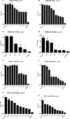
Effect of PQS on cell viability. Human lung epithelial (A549 and NHBE) cells and macrophages (J774A.1 and THP-1) were incubated at 37°C with different concentrations of PQS for 4, 24, and 48 h. Cell viability was assessed at each time point with MTT assays as described in Materials and Methods. Data (percent survival) are the percentages of MTT reduction relative to that of the untreated control and represent the mean ± the SEM of three separate experiments. *, P < 0.05 compared with the control group.
FIG 3
FIG 3
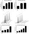
PQS increases ROS production. A549 (A, top), J774A.1 (B, top), NHBE (A, bottom), and THP-1 (B, bottom) cells were cultured in the presence of different concentrations of PQS for 4 or 24 h and then treated with DHE for 30 min as described in Materials and Methods. DHE fluorescence was detected by flow cytometry. Data represent the mean ± the SEM of three separate experiments. *, P < 0.05 compared with the control (non-PQS-treated) group. The inserts above the bar graphs at the bottom show the MFI of cells from the control (C) and columns 1 to 4. DMSO, dimethyl sulfoxide.
FIG 4
FIG 4
PQS increases the fluorescence of ROS-sensitive probes. A549 cells were cultured in the presence of different concentrations of PQS for 4 h and then treated with DCF for 30 min as described in Materials and Methods. DCF fluorescence was detected by confocal microscopy. Control cells show minimal fluorescence (A), and PQS increased DCF (B and C) and DHE (D) fluorescence, indicating increased production of H2O2 and O2˙, respectively. (E) Relative fluorescence intensity per cell of DCF- and DHE-treated cells. Data are the mean ± the SEM of three separate experiments. *, P < 0.05 compared with the control (non-PQS-treated) group. DMSO, dimethyl sulfoxide.
FIG 5
FIG 5
Increased mitochondrial ROS production caused by PQS. J774A.1 macrophages were cultured in the presence of 60 μg/ml PQS for 4 h. Control cells showed minimal fluorescence (A), and PQS increased MitoSOX fluorescence (B). DAPI (blue) stains nuclei in combination with MitoSOX, and cells show increased mitochondrial fluorescence indicating increased production of O2˙ (C). (D) Relative fluorescence intensity per cell of MitoSOX-treated cells. Data represent the mean ± the SEM of three separate experiments. *, P < 0.05 compared with the control (non-PQS-treated) group. DMSO, dimethyl sulfoxide.
FIG 6
FIG 6
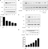
PQS does not change MnSOD or catalase protein expression. A549 lung epithelial cells (A) and J774A.1 macrophages (B) were cultured in the presence of different concentrations of PQS for 24 h. The cells were lysed, and MnSOD, catalase, and actin protein levels were determined by immunoblot analysis as described in Materials and Methods. No significant differences were seen in catalase or MnSOD protein levels in either A549 or J774A.1 cells as a consequence of incubation with PQS. Results are representative of three separate experiments. DMSO, dimethyl sulfoxide.
FIG 7
FIG 7
PQS decreases HO-1 and NrF2 protein expression in lung cells. A549 cells (A), primary NHBE cells (B, C), and macrophages (D) were cultured in the presence of different concentrations of PQS for 24 h. The cells were lysed, and the expression of HO-1 and NrF2 was quantitated by immunoblot analysis as described in Materials and Methods. Expression of HO-1 was significantly decreased in lung epithelial cells (A) and primary bronchial cells (B, C) and increased in J774A.1 macrophage cells by incubation with PQS. NrF2 protein expression was also decreased in both groups of epithelial cells (A to C). PQS exposure also decreased LPS-induced HO-1 expression. Bar graph inserts show results of densitometric analyses of HO-1 blot assays. Data represent the mean ± the SEM of three separate experiments. *, P < 0.05 compared with the control group. DMSO, dimethyl sulfoxide.
FIG 8
FIG 8
PQS induces apoptosis in lung epithelial cells. Shown is early and late apoptosis in A549 (A) and J774A.1 (B) cells following incubation with PQS or the vehicle for 4 h. (A) A dose-dependent increase in the percentage of early, but not late, apoptotic events was seen in PQS-treated A549 cells compared with that of the untreated group. (B) No significant change in early or late apoptosis was seen in J774A.1 cells as a result of exposure to PQS (P > 0.05). The data represent the mean ± the SEM of three independent experiments. *, P < 0.05.

References

    1. Sadikot RT, Blackwell TS, Christman JW, Prince AS. 2005. Pathogen-host interactions in Pseudomonas aeruginosa pneumonia. Am J Respir Crit Care Med 171:1209–1223. doi:10.1164/rccm.200408-1044SO. - DOI - PMC - PubMed
    1. Salins N, Vallath N, Prince V. 2012. Pseudomonas bronchopulmonary infections in a palliative care setting. Indian J Palliat Care 18:1–5. doi:10.4103/0973-1075.97341. - DOI - PMC - PubMed
    1. Delano MJ, Thayer T, Gabrilovich S, Kelly-Scumpia KM, Winfield RD, Scumpia PO, Cuenca AG, Warner E, Wallet SM, Wallet MA, O'Malley KA, Ramphal R, Clare-Salzer M, Efron PA, Mathews CE, Moldawer LL. 2011. Sepsis induces early alterations in innate immunity that impact mortality to secondary infection. J Immunol 186:195–202. doi:10.4049/jimmunol.1002104. - DOI - PMC - PubMed
    1. Lyczak JB, Cannon CL, Pier GB. 2002. Lung infections associated with cystic fibrosis. Clin Microbiol Rev 15:194–222. doi:10.1128/CMR.15.2.194-222.2002. - DOI - PMC - PubMed
    1. Pollack M. 2000. Pseudomonas aeruginosa, p 2310–2335. In Mandell GL, Bennett JE, Dolin R (ed), Principles and practice of infectious diseases, vol 5 Churchill Livingstone, New York, NY.

Publication types

LinkOut - more resources