

Investigation of Seizure-Susceptibility in a Drosophila melanogaster Model of Human Epilepsy with Optogenetic Stimulation
- PMID: 28630111
- PMCID: PMC5560784
- DOI: 10.1534/genetics.116.194779
Investigation of Seizure-Susceptibility in a Drosophila melanogaster Model of Human Epilepsy with Optogenetic Stimulation
Abstract
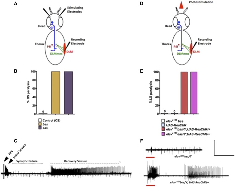
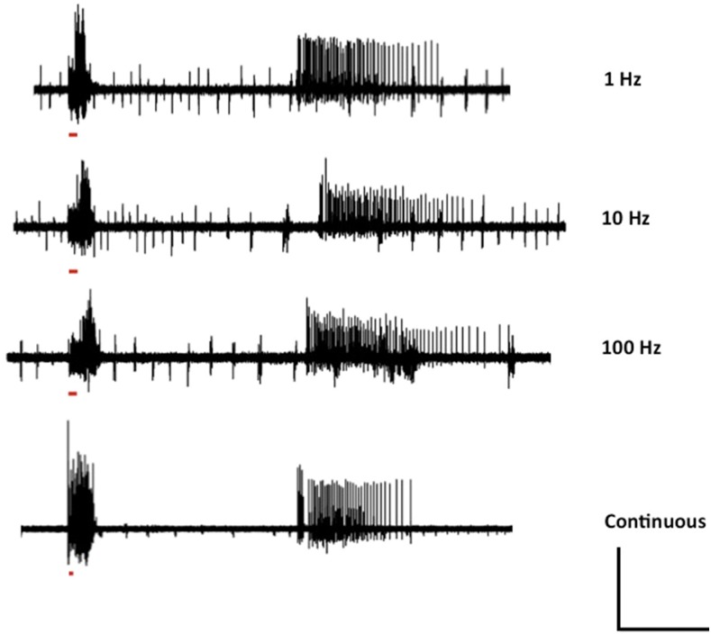
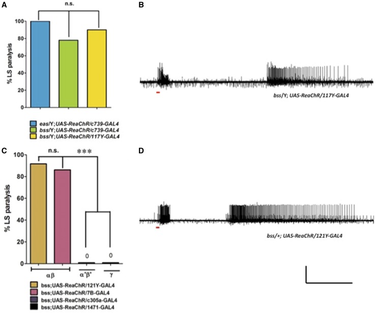
We examined seizure-susceptibility in a Drosophila model of human epilepsy using optogenetic stimulation of ReaChR (red-activatable channelrhodopsin). Photostimulation of the seizure-sensitive mutant parabss1 causes behavioral paralysis that resembles paralysis caused by mechanical stimulation, in many aspects. Electrophysiology shows that photostimulation evokes abnormal seizure-like neuronal firing in parabss1 followed by a quiescent period resembling synaptic failure and apparently responsible for paralysis. The pattern of neuronal activity concludes with seizure-like activity just prior to recovery. We tentatively identify the mushroom body as one apparent locus of optogenetic seizure initiation. The α/β lobes may be primarily responsible for mushroom body seizure induction.
Keywords: epilepsy; red light activable channelrhodopsin; seizure-suppression; sodium channel.
Copyright © 2017 by the Genetics Society of America.
Figures
Similar articles
-
Drosophila as a model for intractable epilepsy: gilgamesh suppresses seizures in para(bss1) heterozygote flies.G3 (Bethesda). 2013 Aug 7;3(8):1399-407. doi: 10.1534/g3.113.006130. G3 (Bethesda). 2013. PMID: 23797108 Free PMC article.
-
Drosophila sodium channel mutations: Contributions to seizure-susceptibility.Exp Neurol. 2015 Dec;274(Pt A):80-7. doi: 10.1016/j.expneurol.2015.06.018. Epub 2015 Jun 18. Exp Neurol. 2015. PMID: 26093037 Free PMC article. Review.
-
Increased persistent Na+ current contributes to seizure in the slamdance bang-sensitive Drosophila mutant.J Neurophysiol. 2011 Jul;106(1):18-29. doi: 10.1152/jn.00808.2010. Epub 2011 Mar 30. J Neurophysiol. 2011. PMID: 21451059 Free PMC article.
-
Seizure control through genetic and pharmacological manipulation of Pumilio in Drosophila: a key component of neuronal homeostasis.Dis Model Mech. 2017 Feb 1;10(2):141-150. doi: 10.1242/dmm.027045. Epub 2016 Dec 14. Dis Model Mech. 2017. PMID: 28067623 Free PMC article.
-
Drosophila melanogaster in the study of epilepsy.SEB Exp Biol Ser. 2008;60:141-60. SEB Exp Biol Ser. 2008. PMID: 18309791 Review. No abstract available.
Cited by
-
The Drosophila ERG channel seizure plays a role in the neuronal homeostatic stress response.PLoS Genet. 2019 Aug 8;15(8):e1008288. doi: 10.1371/journal.pgen.1008288. eCollection 2019 Aug. PLoS Genet. 2019. PMID: 31393878 Free PMC article.
-
Investigating rare and ultrarare epilepsy syndromes with Drosophila models.Fac Rev. 2021 Jan 29;10:10. doi: 10.12703/r/10-10. eCollection 2021. Fac Rev. 2021. PMID: 33659928 Free PMC article. Review.
-
Glia-derived temporal signals orchestrate neurogenesis in the Drosophila mushroom body.Proc Natl Acad Sci U S A. 2021 Jun 8;118(23):e2020098118. doi: 10.1073/pnas.2020098118. Proc Natl Acad Sci U S A. 2021. PMID: 34078666 Free PMC article.
-
Loss-of-function variants in TIAM1 are associated with developmental delay, intellectual disability, and seizures.Am J Hum Genet. 2022 Apr 7;109(4):571-586. doi: 10.1016/j.ajhg.2022.01.020. Epub 2022 Mar 2. Am J Hum Genet. 2022. PMID: 35240055 Free PMC article.
-
Loss of Oxidation Resistance 1, OXR1, Is Associated with an Autosomal-Recessive Neurological Disease with Cerebellar Atrophy and Lysosomal Dysfunction.Am J Hum Genet. 2019 Dec 5;105(6):1237-1253. doi: 10.1016/j.ajhg.2019.11.002. Epub 2019 Nov 27. Am J Hum Genet. 2019. PMID: 31785787 Free PMC article.
References
Publication types
MeSH terms
Substances
Grants and funding
LinkOut - more resources
Full Text Sources
Other Literature Sources
Medical
Molecular Biology Databases
