. 2017 Aug 8;199(17):e00101-17.
doi: 10.1128/JB.00101-17. Print 2017 Sep 1.

Absence of the Polar Organizing Protein PopZ Results in Reduced and Asymmetric Cell Division in Agrobacterium tumefaciens

Affiliations

Absence of the Polar Organizing Protein PopZ Results in Reduced and Asymmetric Cell Division in Agrobacterium tumefaciens

Matthew Howell et al. J Bacteriol. .

Abstract

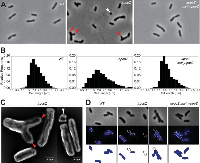
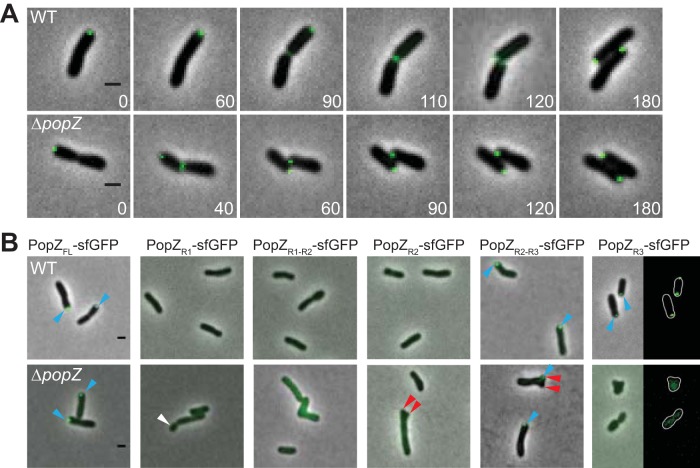
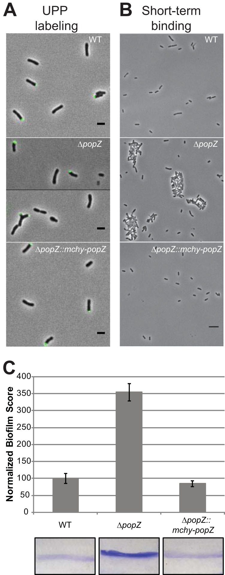
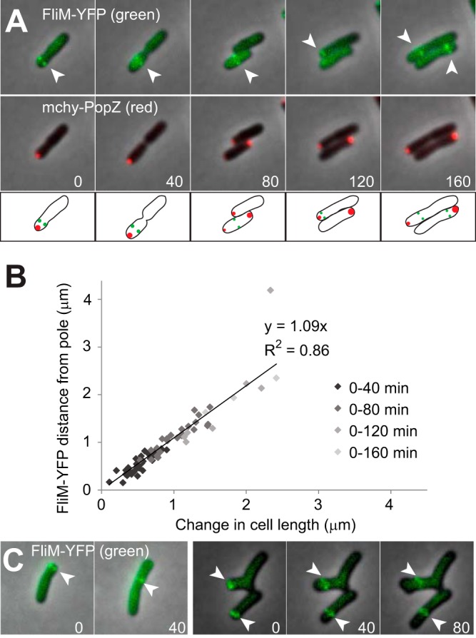
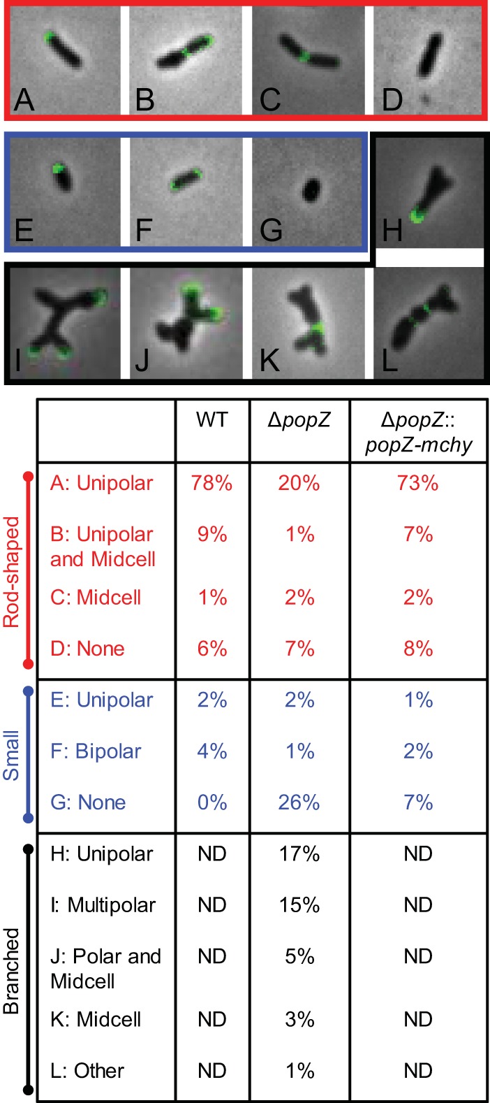
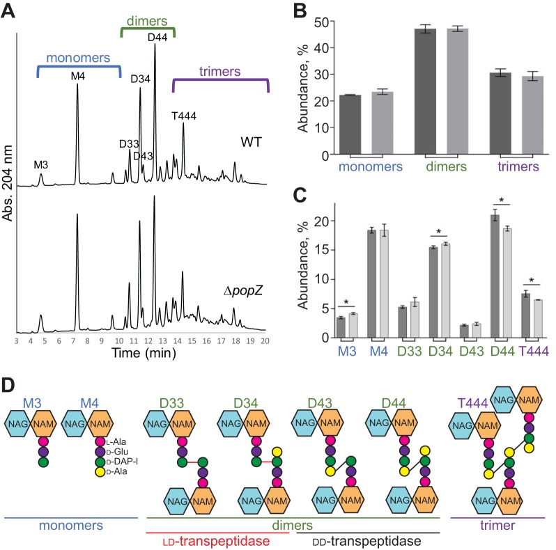
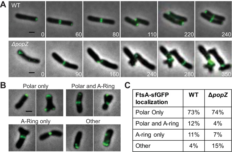
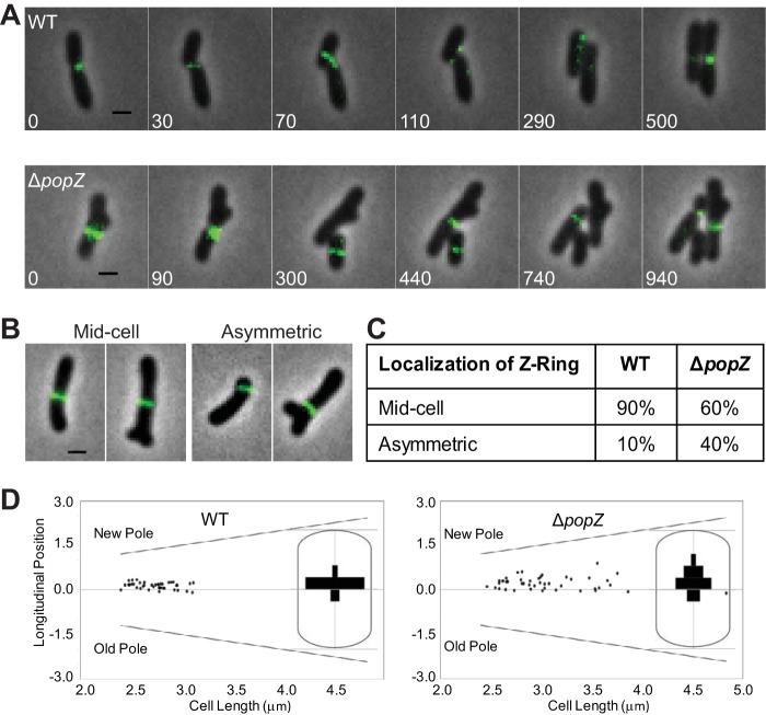
Agrobacterium tumefaciens is a rod-shaped bacterium that grows by polar insertion of new peptidoglycan during cell elongation. As the cell cycle progresses, peptidoglycan synthesis at the pole ceases prior to insertion of new peptidoglycan at midcell to enable cell division. The A. tumefaciens homolog of the Caulobacter crescentus polar organelle development protein PopZ has been identified as a growth pole marker and a candidate polar growth-promoting factor. Here, we characterize the function of PopZ in cell growth and division of A. tumefaciens Consistent with previous observations, we observe that PopZ localizes specifically to the growth pole in wild-type cells. Despite the striking localization pattern of PopZ, we find the absence of the protein does not impair polar elongation or cause major changes in the peptidoglycan composition. Instead, we observe an atypical cell length distribution, including minicells, elongated cells, and cells with ectopic poles. Most minicells lack DNA, suggesting a defect in chromosome segregation. Furthermore, the canonical cell division proteins FtsZ and FtsA are misplaced, leading to asymmetric sites of cell constriction. Together, these data suggest that PopZ plays an important role in the regulation of chromosome segregation and cell division.IMPORTANCEA. tumefaciens is a bacterial plant pathogen and a natural genetic engineer. However, very little is known about the spatial and temporal regulation of cell wall biogenesis that leads to polar growth in this bacterium. Understanding the molecular basis of A. tumefaciens growth may allow for the development of innovations to prevent disease or to promote growth during biotechnology applications. Finally, since many closely related plant and animal pathogens exhibit polar growth, discoveries in A. tumefaciens may be broadly applicable for devising antimicrobial strategies.

Keywords: Agrobacterium; PopZ; cell division; cell polarity; chromosome segregation; growth polarity.


Figures

FIG 1
FIG 1
Analysis of morphology, cell length, and DNA content of the popZ deletion strain. (A) Comparison of phase-contrast images of wild-type, ΔpopZ, and ΔpopZ::popZ-mchy strains grown to exponential phase in ATGN media. The ΔpopZ culture contains a high proportion of small cells (<1.5 μm in length; white arrowhead) and branched cells with ectopic poles (red arrowheads). (B) Cell length distributions of WT (left; n = 926), ΔpopZ (center; n = 1,664), and ΔpopZ::popZ-mchy (n = 839) cells are shown. (C) Transmission electron micrographs of nano-tungsten-stained ΔpopZ cells. The deletion of popZ results in an increased cell length distribution, including very small cells (white arrowhead) and cells with ectopic poles (red arrowheads). (D) DAPI staining reveals the presence of anucleate cells in the ΔpopZ population. Phase (top) and fluorescent (middle) images of representative DAPI-stained wild-type, ΔpopZ, and ΔpopZ::popZ-mchy cells are shown. Outlines are provided to indicate cell location in fluorescent images. Schematics of DAPI-stained cells are provided in the bottom panel.
FIG 2
FIG 2
Polar localization of PopZ requires domains R2 and R3. (A, top) Time-lapse microscopy of WT cells expressing full-length PopZ-sfGFP (PopZFL-sfGFP) reveals a new pole-to-midcell localization pattern. (Bottom) PopZFL-sfGFP exhibits a similar localization pattern in the ΔpopZ strain. (B) Representative images of the localization patterns of full-length and truncated versions of PopZ-sfGFP in wild-type and ΔpopZ cells are shown. In wild-type cells, PopZ truncations containing domain R3 (PopZFL-sfGFP, PopZR2-R3-sfGFP, and PopZR3-sfGFP) localize to growth poles (blue arrowheads). In the absence of PopZ, only PopZFL-sfGFP and PopZR2-R3-sfGFP localize to growth poles (blue arrowheads). Truncated forms of PopZ do not complement the morphological defects of ΔpopZ cells, as indicated by the presence of small cells (white arrowheads) and ectopic poles (red arrowheads). All scale bars are 1 μm.
FIG 3
FIG 3
Analysis of biofilm formation in wild-type and ΔpopZ mutant strains. (A) UPP production and placement was identified by the binding of Alexa Fluor 488-labeled wheat germ agglutinin to cells on 1.5% agarose pads. In all three strains, approximately 20% of individual cells have a detectable UPP. Scale bars, 2 μm. (B) Short-term binding was evaluated after 1 h of attachment to glass coverslips. Scale bar, 5 μm. (C) Strains were assayed for biofilm formation on vertical plastic coverslips immersed in ATGN medium. Coverslips were removed after 48 h of incubation at room temperature and rinsed to remove any loosely associated cells. Adherent biomass was determined as the absorbance of solubilized crystal violet (A600), and the optical density of the planktonic culture (OD600) was measured. Biofilm scores were calculated as the A600/OD600 ratio, and data were normalized to WT values. Data shown are the means from two independent experiments completed in triplicate. Error bars are the standard errors of the means. Representative coverslips prior to crystal violet solubilization are shown for each strain.
FIG 4
FIG 4
Analysis of flagellum localization in wild-type and ΔpopZ mutant strains. (A) Flagellar basal bodies were fluorescently labeled by expressing FliM-YFP, and their localization patterns (green) were observed by time-lapse fluorescence microscopy in cells that also express mChy-PopZ (red). FliM-YFP foci are indicated by arrowheads. Fluorescence images are overlaid on a phase-contrast background, and time (in minutes) is shown in the lower corners of images. Schematics are provided below the images. (B) For cells shown in panel A, a scatterplot compares the increase in cell length to the increase in the distance of the FliM-YFP foci from the cell poles. A total of 34 cells were measured in 2 independent experiments, and the slope of the linear regression and associated R-squared value is shown. There are fewer data points for later time points because most cells divided during the time course. (C) Fluorescence localization of FliM-YFP (green) in ΔpopZ cells.
FIG 5
FIG 5
Deletion of popZ causes atypical growth patterning. Fluorescent d-amino-acid labeling was used as a proxy for sites of peptidoglycan synthesis in WT, ΔpopZ, and ΔpopZ::popZ-mchy strains. (Top) Representative images of cellular morphology and growth patterns from the ΔpopZ mutant are shown. Patterns observed in typical rod-shaped bacteria (red), small cells (<1.5 μm; blue), and branched cells (black) are shown and quantitated in the table below. One hundred fifty cells were labeled and categorized as shown in the table for each strain. Branched cells were not detected (ND) in WT and ΔpopZ::popZ-mchy strains.
FIG 6
FIG 6
Peptidoglycan composition is slightly modified in ΔpopZ cells. (A) UPLC spectra of muropeptides derived from WT or ΔpopZ cells. Major muropeptides are labeled. M, monomers; D, dimers; T, trimers. Numbers indicate the length of the muropeptide stems and the position of cross-links in dimers and trimers. (B) Abundance of total monomers, dimers, and trimers in the muropeptide profile for WT (dark gray) and ΔpopZ (light gray) cells. (C) Quantitation of the major muropeptide peaks in WT (dark gray) and ΔpopZ (light gray) cells. For panels C and D, data shown are the average abundances of each muropeptide and are taken from analysis of three independent biological samples. Statistical significance is indicated with an asterisk. (D) Schematics of major muropeptides are shown. The monomers (blue), dimers (green), and trimer (purple) are labeled. Note that the type of cross-link is shown for each dimer and trimer. dd-Cross-links are shown in black, and ld-cross-links are shown in red.
FIG 7
FIG 7
FtsA forms polar foci and rings in ΔpopZ cells. (A) Time-lapse microscopy of FtsA-sfGFP in WT cells (top) shows a polar-to-midcell localization pattern. Time-lapse microscopy of FtsA-sfGFP in the ΔpopZ mutant (bottom) shows cells with unipolar foci, multipolar foci, and both stable and unstable ring structures. (B) Representative examples of FtsA-sfGFP localization patterns observed in ΔpopZ cells. All scale bars are 1 μm. (C) Percentage of cells with each category of FtsA-sfGFP localization pattern in WT (309 cells) and ΔpopZ (328 cells) strains.
FIG 8
FIG 8
Analysis of FtsZ localization and constriction sites in ΔpopZ cells. (A, top) Time-lapse microscopy of FtsZ-sfGFP in WT cells shows dynamic localization of FtsZ foci before Z-ring formation at midcell. (Bottom) Time-lapse microscopy of FtsZ-sfGFP in ΔpopZ cells reveals atypical Z-ring size and positioning. (B) Representative images showing midcell and asymmetric Z-ring of ΔpopZ cells. All scale bars are 1 μm. (C) Quantification of Z-ring position in WT and ΔpopZ cells. Z-rings greater than 0.15 μm from midcell in either direction are defined as asymmetric. For this analysis, 77 Z-rings were analyzed in WT cells and 60 Z-rings were analyzed in ΔpopZ cells. (D) The longitudinal position of constriction sites is plotted against the cell length for 41 WT and 41 ΔpopZ cells. The position of each pole is shown with a diagonal line. A longitudinal position of zero is midcell. Positive values are closer to the new pole, whereas negative values are closer to the old pole. Insets show histograms illustrating the longitudinal position of constriction sites within a cell.

Similar articles

Cited by

References

    1. Brown PJB, de Pedro MA, Kysela DT, Van der Henst C, Kim J, De Bolle X, Fuqua C, Brun YV. 2012. Polar growth in the alphaproteobacterial order Rhizobiales. Proc Natl Acad Sci U S A 109:1697–1701. doi:10.1073/pnas.1114476109. - DOI - PMC - PubMed
    1. Kuru E, Velocity Hughes H, Brown PJ, Hall E, Tekkam S, Cava F, de Pedro MA, Brun YV, VanNieuwenhze MS. 2012. In situ probing of newly synthesized peptidoglycan in live bacteria with fluorescent D-amino acids. Angew Chem Int Ed Engl 51:12519–12523. doi:10.1002/anie.201206749. - DOI - PMC - PubMed
    1. Cameron TA, Anderson-Furgeson J, Zupan JR, Zik JJ, Zambryski PC. 2014. Peptidoglycan synthesis machinery in Agrobacterium tumefaciens during unipolar growth and cell division. mBio 5:e01219-14. doi:10.1128/mBio.01219-14. - DOI - PMC - PubMed
    1. Margolin W. 2009. Sculpting the bacterial cell. Curr Biol 19:R812–R822. doi:10.1016/j.cub.2009.06.033. - DOI - PMC - PubMed
    1. Grangeon R, Zupan JR, Anderson-Furgeson J, Zambryski PC. 2015. PopZ identifies the new pole, and PodJ identifies the old pole during polar growth in Agrobacterium tumefaciens. Proc Natl Acad Sci U S A 112:11666–11671. doi:10.1073/pnas.1515544112. - DOI - PMC - PubMed

MeSH terms

LinkOut - more resources