. 2017 Sep;23(9):1465-1476.
doi: 10.1261/rna.060632.117. Epub 2017 Jun 19.

Three-dimensional structure of the 3'X-tail of hepatitis C virus RNA in monomeric and dimeric states

Affiliations

Three-dimensional structure of the 3'X-tail of hepatitis C virus RNA in monomeric and dimeric states

Ángel Cantero-Camacho et al. RNA. 2017 Sep.

Abstract

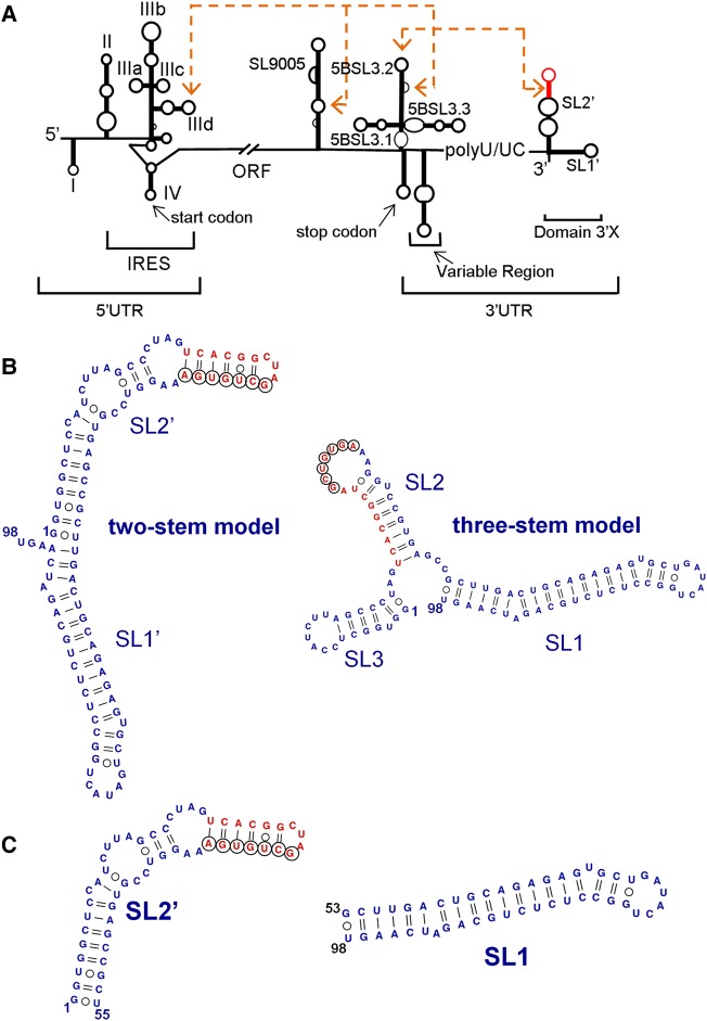
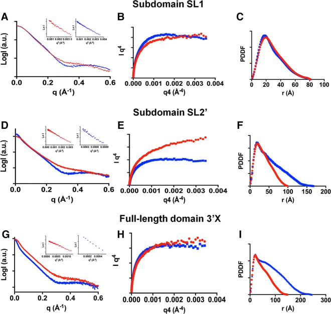
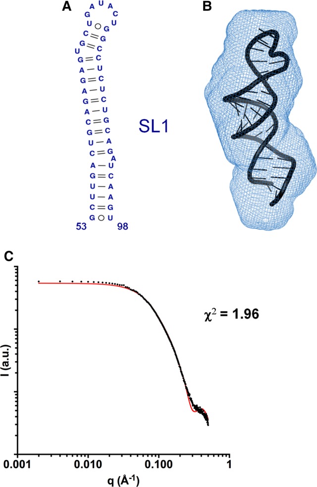
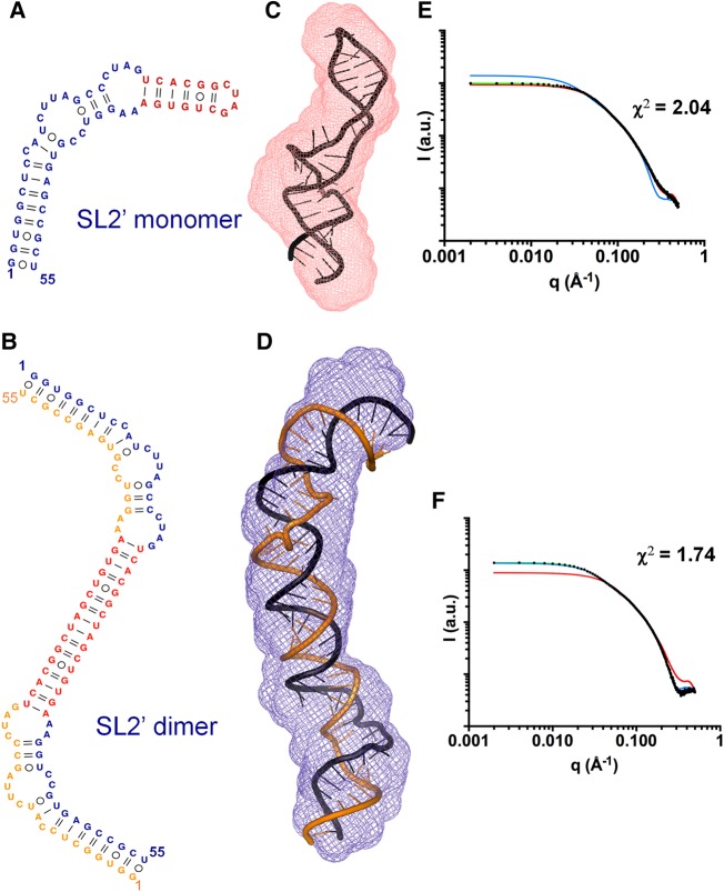
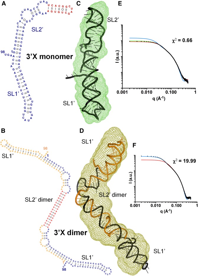
The 3'X domain is a 98-nt region located at the 3' end of hepatitis C virus genomic RNA that plays essential functions in the viral life cycle. It contains an absolutely conserved, 16-base palindromic sequence that promotes viral RNA dimerization, overlapped with a 7-nt tract implicated in a distal contact with a nearby functional sequence. Using small angle X-ray scattering measurements combined with model building guided by NMR spectroscopy, we have studied the stoichiometry, structure, and flexibility of domain 3'X and two smaller subdomain sequences as a function of ionic strength, and obtained a three-dimensional view of the full-length domain in its monomeric and dimeric states. In the monomeric form, the 3'X domain adopted an elongated conformation containing two SL1' and SL2' double-helical stems stabilized by coaxial stacking. This structure was significantly less flexible than that of isolated subdomain SL2' monomers. At higher ionic strength, the 3'X scattering envelope nearly doubled its size, reflecting the formation of extended homodimers containing an antiparallel SL2' duplex flanked by coaxially stacked SL1' helices. Formation of these dimers could initialize and/or regulate the packaging of viral RNA genomes into virions.

Keywords: 3′X domain; RNA; dimer; hepatitis C virus; small-angle X-ray scattering; structure.


Figures

FIGURE 1.
FIGURE 1.
HCV RNA and 3′X terminal domain. (A) Functional structures identified in the HCV genomic RNA; distal RNA–RNA interactions proposed to have functional relevance are identified. (B) Secondary structure of two alternative conformations of full-length domain 3′X. These structures expose differently the absolutely conserved, overlapping sequences k (indicated with black circles) and DLS (red nt). Sequence k has been proposed to establish a distal kissing-loop interaction with a complementary k′ sequence located in the apical loop of ORF hairpin 5BSL3.2, and DLS is a 16-nt palindromic sequence likely involved in HCV RNA dimerization. (C) Secondary structure of 3′X subdomains SL2′ (also identified as X55 in the literature) and SL1.
FIGURE 2.
FIGURE 2.
Small-angle X-ray scattering data of subdomains SL1 and SL2′ and full-length domain 3′X of hepatitis C virus RNA. (A,D,G) Scattering intensities versus momentum transfer q, obtained at low (red) and higher (blue) ionic strength. The insets show, for each ionic strength condition, the Guinier region of the scattering curve with a linear fit line. (B,E,H) Porod–Debye plots at low (red) and higher (blue) ionic strength. (C,F,I) Pair distance distribution functions (PDDF) at low (red) and higher (blue) ionic strength. Experimental conditions: 0.5–2 mg mL−1 (subdomains SL1 and SL2′) or 0.375–1.5 mg mL−1 (full-length domain 3′X); 10 mM Tris–HCl pH 7.0, 0 mM NaCl/MgCl2 (all sequences, low ionic strength); 10 mM Tris–HCl pH 7.0 and 2 mM MgCl2 (subdomains SL1 and SL2′, higher ionic strength); or 10 mM Tris–HCl pH 7.0, 2 mM MgCl2 and 50 mM NaCl (domain 3′X, higher ionic strength).
FIGURE 3.
FIGURE 3.
Three-dimensional structure of subdomain SL1 determined by SAXS. (A) Secondary structure of SL1 detected by NMR spectroscopy at low and higher salt concentration (Cantero-Camacho and Gallego 2015). (B) Average ab initio envelope of SL1 calculated from the SAXS profile at low ionic strength, superposed with the best energy-minimized SL1 atomic model, selected by fitting the theoretical SAXS profiles to the experimental curve. The normalized spatial discrepancy (NSD) between scattering envelopes was 0.624. (C) Theoretical SAXS profile calculated from the best energy-minimized SL1 atomic model (red line), overlaid with the experimental profile obtained at low ionic concentration (black dots).
FIGURE 4.
FIGURE 4.
SAXS structural analysis of subdomain SL2′. (A,B) Secondary structures of SL2′ determined by NMR spectroscopy. This sequence formed monomeric hairpins at low ionic strength (A) and symmetric dimers in the presence of salts (B) (Cantero-Camacho and Gallego 2015). (C,D) Average ab initio envelopes of SL2′ obtained by SAXS analyses in conditions of low (C) and higher (D) ionic strength, superposed with the best energy-minimized respective atomic models, selected by fitting the theoretical scattering profiles to the experimental curves. The NSD values between SAXS envelopes were 0.560 and 0.555, respectively. (E,F) Experimental SAXS curves (black dots) of subdomain SL2′ obtained at low (E) and higher (F) salt concentration, overlaid with the theoretical profiles calculated from the best energy-minimized monomer (red lines) and dimer (blue lines) models, and from a mixture of monomer and dimer models (green lines).
FIGURE 5.
FIGURE 5.
Solution structure of full-length domain 3′X of HCV RNA. (A,B) Secondary structures of domain 3′X supported by NMR experiments carried out under conditions of low (A) and higher (B) ionic strength. This sequence was found to form monomers or symmetric dimers under these respective solution conditions (Cantero-Camacho and Gallego 2015). (C,D) Average ab initio envelopes of domain 3′X obtained from the SAXS profiles at low (C) and higher (D) salt concentration, superposed with the best energy-minimized respective atomic models, selected by fitting the computed SAXS profiles to the experimental curves. The NSDs were 0.573 and 0.783, respectively. (E,F) Experimental SAXS profiles of domain 3′X obtained at low (E) and higher (F) salt concentration (black dots), overlaid with the theoretical curves calculated from the best energy-minimized monomer (red lines) and dimer (blue lines) models, and from a mixture of monomer and dimer models (green lines). In F, the relatively higher χ2 value is due to a smaller experimental error of the measured curve (see χ-function in Schneidman-Duhovny et al. 2010).

Similar articles

Cited by

References

    1. Ali M, Lipfert J, Seifert S, Herschlag D, Doniach S. 2010. The ligand-free state of the TPP riboswitch: a partially folded RNA structure. J Mol Biol 396: 153–165. - PMC - PubMed
    1. Bartenschlager R, ed. 2013. Hepatitis C virus: from molecular virology to antiviral therapy. Springer-Verlag, Berlin.
    1. Blight KJ, Rice CM. 1997. Secondary structure determination of the conserved 98-base sequence at the 3′ terminus of hepatitis C virus genome RNA. J Virol 71: 7345–7352. - PMC - PubMed
    1. Burke JE, Butcher SE. 2012. Nucleic acid structure characterization by small angle X-ray scattering (SAXS). Curr Protoc Nucleic Acid Chem 51: 7.18.1–7.18.18. - PMC - PubMed
    1. Cai Z, Liang TJ, Luo G. 2004. Effects of mutations of the initiation nucleotides on hepatitis C virus RNA replication in the cell. J Virol 78: 3633–3643. - PMC - PubMed

Publication types

LinkOut - more resources