Review
. 2017 Aug 5;372(1726):20160209.
doi: 10.1098/rstb.2016.0209.

Molecular mechanism of pore formation by aerolysin-like proteins

Affiliations
Review

Molecular mechanism of pore formation by aerolysin-like proteins

Marjetka Podobnik et al. Philos Trans R Soc Lond B Biol Sci. .

Abstract

Aerolysin-like pore-forming proteins are an important family of proteins able to efficiently damage membranes of target cells by forming transmembrane pores. They are characterized by a unique domain organization and mechanism of action that involves extensive conformational rearrangements. Although structures of soluble forms of many different members of this family are well understood, the structures of pores and their mechanism of assembly have been described only recently. The pores are characterized by well-defined β-barrels, which are devoid of any vestibular regions commonly found in other protein pores. Many members of this family are bacterial toxins; therefore, structural details of their transmembrane pores, as well as the mechanism of pore formation, are an important base for future drug design. Stability of pores and other properties, such as specificity for some cell surface molecules, make this family of proteins a useful set of molecular tools for molecular recognition and sensing in cell biology.This article is part of the themed issue 'Membrane pores: from structure and assembly, to medicine and technology'.

Keywords: aerolysin; lysenin; membranes; monalysin; pore formation; pore-forming protein.


Conflict of interest statement

We have no competing interests.

Figures

Figure 1.
Figure 1.
Structures of some of aβ-PFPs representatives. (a) Ribbon representation of some of the most studied representatives of aβ-PFPs, and their PDB-IDs are in the brackets below their names. RBDs are shown in grey and PFMs as: blue, conserved core; red, a variable loop; orange, a weakly conserved β-strand; cyan, an insertion loop. (b) Topological diagram of the PFM of three representatives of aβ-PFPs, colouring the same as in (a). β-Strands are numbered and coloured according to the consensus from Szczesny et al. [5].
Figure 2.
Figure 2.
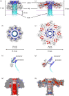
Structural features of aβ-PFPs pores. (ad) Lysenin pore (PDB-ID 5EC5). (a) Ribbon presentation of the lysenin pore, side view and (b) top view. Pore dimensions are shown, as well as membrane position (pink lines). (c) Superposition of the lysenin monomer (PDB-ID 3XZD) and a protomer from a pore, superposition on the C-terminal domain. (d) The electrostatic properties of the inner surface of the model of the wild-type lysenin pore [8]. The surface of the pore is coloured according to electrostatic potential. A cut-off of −5 kT e−1 was used for the negative potential (red) and of +5 kT e−1 for the positive potential (blue). (e–h) Aerolysin pore (PDB-ID 5JZT). (e) Ribbon presentation of the aerolysin pore, side view and (f) top view. Pore dimensions as well as membrane position (lines) are shown. (g) Superposition of the pro-aerolysin monomer (PDB-ID 1PRE) and a protomer from a pore, superposition on helices of domain D2. (h) The electrostatic properties of the inner surface of the model of the aerolysin pore [9]. The surface of the pore is coloured as in (d).
Figure 3.
Figure 3.
Mechanism of pore assembly by aβ-PFPs based on the structure of the lysenin monomer and its pore. Step 1: Soluble proteins (i.e. monomeric, dimeric) bind to their respective receptors (green circles/sticks for lysenin receptor sphingomyelin) on the membrane (pink) with their RBD domain (grey circle). Dimers of aerolysin or Dln1 dissociate upon binding. aβ-PFPs like monalysin do not require any specific receptor but instead form large soluble complexes in order to increase their concentration at the membrane. Step 2: Bound monomers start oligomerizing above the membrane to form ring-shaped prepores. Many aβ-PFPs contain propeptides, which need to be removed to initiate oligomerization. It has been shown for lysenin that the prepore grows via gradual elongation of membrane-bound arcs. The shape of the prepore depends on the subdomain structure of aβ-PFP, being cone-like for aerolysin and wreath-like for lysenin, resulting in heptameric and nonameric prepore, respectively. In the case of monalysin, the nonameric prepore seems to be already included in the doughnut-shaped soluble form that dissociates upon propeptide cleavage, resulting in nonameric disks landing on a membrane as prepores. Transition from the monomer structure to prepore can already require some conformational changes, as in the case of aerolysin, which seems not to be needed in lysenin. Step 3: Finally, large conformational changes in PFM domain enable insertion of approximately half of this domain into the membrane and formation of the pore with β-barrel channel. PFM is built of two parts (cyan for insertion loop and orange/blue for the twisted β-sheet that does not change its intrinsic secondary structure upon pore formation). Colours used are the same as in figure 1. In steps 2 and 3, only three protomers of the oligomer are shown for clarity. On average, the number of protomers in ring-like prepores and pores ranges between six and nine, depending on the protein involved.

Similar articles

Cited by

References

    1. Anderluh G, Lakey JH. 2008. Disparate proteins use similar architectures to damage membranes. Trends Biochem. Sci. 33, 482–490. (10.1016/j.tibs.2008.07.004) - DOI - PubMed
    1. Gilbert RJ, Dalla Serra M, Froelich CJ, Wallace MI, Anderluh G. 2014. Membrane pore formation at protein–lipid interfaces. Trends Biochem. Sci. 39, 510–516. (10.1016/j.tibs.2014.09.002) - DOI - PubMed
    1. Sugawara T, Yamashita D, Kato K, Peng Z, Ueda J, Kaneko J, Kamio Y, Tanaka Y, Yao M. 2015. Structural basis for pore-forming mechanism of staphylococcal alpha-hemolysin. Toxicon 108, 226–231. (10.1016/j.toxicon.2015.09.033) - DOI - PubMed
    1. Anderluh G, Gilbert RJ (eds) 2014. MACPF/CDC proteins—agents of defence, attack and invasion. The Netherlands: Springer.
    1. Szczesny P, Iacovache I, Muszewska A, Ginalski K, van der Goot FG, Grynberg M.. 2011. Extending the aerolysin family: from bacteria to vertebrates. PLoS ONE 6, e20349 (10.1371/journal.pone.0020349) - DOI - PMC - PubMed

MeSH terms

LinkOut - more resources