Review
. 2017 Aug 5;372(1726):20160217.
doi: 10.1098/rstb.2016.0217.

Apoptotic foci at mitochondria: in and around Bax pores

Affiliations
Review

Apoptotic foci at mitochondria: in and around Bax pores

Begoña Ugarte-Uribe et al. Philos Trans R Soc Lond B Biol Sci. .

Abstract

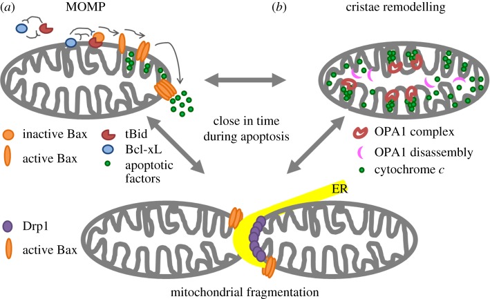
The permeabilization of the mitochondrial outer membrane by Bax and Bak during apoptosis is considered a key step and a point of no return in the signalling pathway. It is always closely related to the reorganization of mitochondrial cristae that frees cytochrome c to the intermembrane space and to massive mitochondrial fragmentation mediated by the dynamin-like protein Drp1. Despite multiple evidence in favour of a functional link between these processes, the molecular mechanisms that connect them and their relevance for efficient apoptosis signalling remain obscure. In this review, we discuss recent progress on our understanding of how Bax forms pores in the context of Drp1-stabilized signalling platforms at apoptotic foci in mitochondria.This article is part of the themed issue 'Membrane pores: from structure and assembly, to medicine and technology'.

Keywords: Bcl-2; MOMP; cristae remodelling; membrane curvature; mitochondrial fragmentation.


Conflict of interest statement

We declare we have no competing interests.

Figures

Figure 1.
Figure 1.
Scheme of structural changes in mitochondria during apoptosis: MOMP (a), cristae remodelling (b), mitochondrial fragmentation (bottom).
Figure 2.
Figure 2.
Bax pore architecture. (a) Magnified reconstructed super-resolution images of Bax pores in apoptotic HeLa cells corresponding to arc (left) and ring (right) structures (with permission from [95]). SMLM stands for Single Molecule Localization Microscopy. Scale bars, 100 nm. (b) Three-dimensional atomic force microscopy (AFM) topography of a Bax structure in a supported lipid bilayer (right) and its height profile (left) corresponding to the pore rim. (c) Representative scheme of Bax toroidal pore (top view) where both Bax dimers and lipids cooperate for pore formation. The structure of active, membrane-inserted Bax dimer is used based on the ‘clamp’ model [100].
Figure 3.
Figure 3.
Role of Drp1 in mitochondrial fission and apoptosis. (a) Drp1 shuttles between cytosol and mitochondria for membrane division under physiological conditions. Overview (top panel) and zoomed image of the white box (bottom panel). Immunostaining of endogenous Drp1 (green) and mitochondrial staining with Mitotracker Red (magenta). Scale bar, 5 µm. (b) Drp1 (green) and Bax (magenta) binding to giant unilamellar vesicles composed of phosphatidylcholine: phosphatidylethanolamine: cardiolipin (54 : 20 : 26, mol : mol) at 42° for 30 min. Drp1 and Bax are concentrated at contact surfaces between two vesicles. Bax clusters at highly curved edges between vesicles. Scale bar, 10 µm.

Similar articles

Cited by

References

    1. Martinou JC, Youle RJ. 2011. Mitochondria in apoptosis: Bcl-2 family members and mitochondrial dynamics. Dev. Cell 21, 92–101. (10.1016/j.devcel.2011.06.017) - DOI - PMC - PubMed
    1. Osman C, Voelker DR, Langer T. 2011. Making heads or tails of phospholipids in mitochondria. J. Cell Biol. 192, 7–16. (10.1083/jcb.201006159) - DOI - PMC - PubMed
    1. Glancy B, Balaban RS. 2012. Role of mitochondrial Ca2+ in the regulation of cellular energetics. Biochemistry 51, 2959–2973. (10.1021/bi2018909) - DOI - PMC - PubMed
    1. Ugarte-Uribe B, García-Sáez AJ. 2014. Membranes in motion: mitochondrial dynamics and their role in apoptosis. Biol. Chem. 395, 297–311. (10.1515/hsz-2013-0234) - DOI - PubMed
    1. Horvath SE, Daum G. 2013. Lipids of mitochondria. Prog. Lipid Res. 52, 590–614. (10.1016/j.plipres.2013.07.002) - DOI - PubMed

Substances

LinkOut - more resources