Review
. 2017 Aug 5;372(1726):20160218.
doi: 10.1098/rstb.2016.0218.

Pore formation by dimeric Bak and Bax: an unusual pore?

Affiliations
Review

Pore formation by dimeric Bak and Bax: an unusual pore?

Rachel T Uren et al. Philos Trans R Soc Lond B Biol Sci. .

Abstract

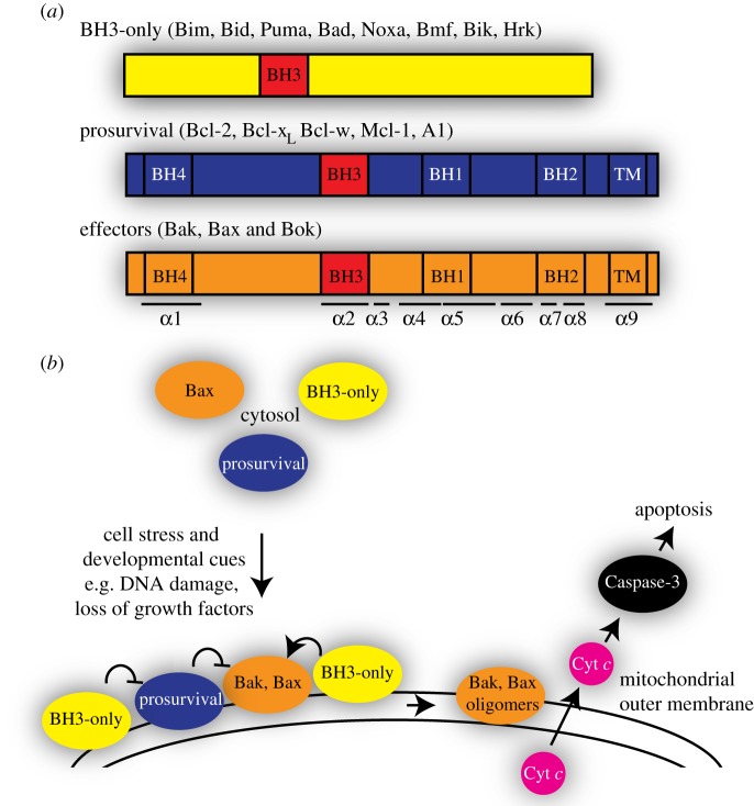
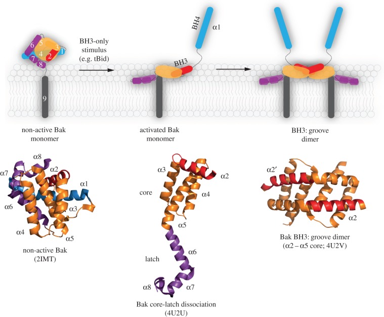
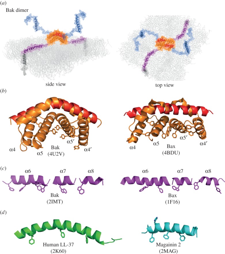
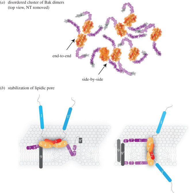
Apoptotic cell death via the mitochondrial pathway occurs in all vertebrate cells and requires the formation of pores in the mitochondrial outer membrane. Two Bcl-2 protein family members, Bak and Bax, form these pores during apoptosis, and how they do so has been investigated for the last two decades. Many of the conformation changes that occur during their transition to pore-forming proteins have now been delineated. Notably, biochemical, biophysical and structural studies indicate that symmetric homodimers are the basic unit of pore formation. Each dimer contains an extended hydrophobic surface that lies on the outer membrane, and is anchored at either end by a transmembrane domain. Membrane-remodelling events such as positive membrane curvature have been reported to accompany apoptotic pore formation, suggesting Bak and Bax form lipidic pores rather than proteinaceous pores. However, it remains unclear how symmetric dimers assemble to porate the membrane. Here, we review how clusters of dimers and their lipid-mediated interactions provide a molecular explanation for the heterogeneous assemblies of Bak and Bax observed during apoptosis.This article is part of the themed issue 'Membrane pores: from structure and assembly, to medicine and technology'.

Keywords: Bak; Bax; apoptosis; heterogeneity; membrane bilayer; mitochondrial pore.


Conflict of interest statement

We declare we have no competing interests.

Figures

Figure 1.
Figure 1.
Bcl-2 proteins regulate the mitochondrial pathway of apoptotic cell death. (a) Three subfamilies of Bcl-2 proteins. (b) Bak and Bax activation by the BH3-only proteins is followed by their oligomerization in the mitochondrial outer membrane to release cytochrome c and induce apoptosis.
Figure 2.
Figure 2.
Bak activation and conformation change results in symmetric homodimers. A schematic showing that Bak unfolds by the N-terminus (α1, blue) and the C-terminal latch (α6–α8, magenta) separating from the α2–α5 core (orange, red). Hydrophobic regions of the core and latch then collapse onto the membrane, while the exposed BH3 domain (in α2) binds to the hydrophobic groove in another activated Bak molecule. Reciprocal BH3:groove binding results in symmetric homodimers. The indicated crystal structures demonstrate the major conformation changes involved. Equivalent changes are observed for Bax.
Figure 3.
Figure 3.
Membrane topology of the Bak dimer. (a) The in-plane model of the Bak dimer. The N-terminal regions become solvent-exposed while the remainder of the Bak dimer resembles a flexible extended amphipathic peptide that lies in-plane with the membrane, anchored at either end by transmembrane domains. Note that α1 may unfold after it dissociates, decreasing the hydrophobicity of the BH4 structural motif (VFrsYV) therein [10,21]. Images were assembled in PyMol using the structures of Bak (2IMT) and the Bak dimer (4U2V), and represented as cartoon and mesh. (b) Aromatic residues are concentrated on the bent surface of the Bak and Bax α2–α5 core dimers. (c) Aromatic residues can position on one edge of the flexible α6–α8 latch. (d) Examples of antimicrobial peptides thought to form lipidic pores, with aromatic residues indicated. Colour coding as in figure 2.
Figure 4.
Figure 4.
Possible mechanisms involved in lipidic pore formation and stabilization by homodimers of Bak or Bax. (a) Schematic of Bak dimers forming a disordered cluster on the mitochondrial outer membrane, encouraged by flexibility of the α6–α8 latch. Note that end-to-end or side-by-side contact between the core regions is possible. Images were assembled as in figure 3a. (b) Parts of the dimer may line a lipidic pore. The flexible amphipathic latch may slide into a nascent pore to partially line and stabilize the pore (left; in-plane model [4]). The amphipathic core dimer (α2–α5) may also line the pore generating antiparallel α9-helices (right; clamp model [19]). Colour coding as in figure 2.

References

    1. Czabotar PE, Lessene G, Strasser A, Adams JM. 2014. Control of apoptosis by the BCL-2 protein family: implications for physiology and therapy. Nat. Rev. Mol. Cell Biol. 15, 49–63. ( 10.1038/nrm3722) - DOI - PubMed
    1. Lindsten T, et al. 2000. The combined functions of proapoptotic Bcl-2 family members Bak and Bax are essential for normal development of multiple tissues. Mol. Cell 6, 1389–1399. ( 10.1016/S1097-2765(00)00136-2) - DOI - PMC - PubMed
    1. Wei MC, et al. 2001. Proapoptotic BAX and BAK: a requisite gateway to mitochondrial dysfunction and death. Science 292, 727–730. ( 10.1126/science.1059108) - DOI - PMC - PubMed
    1. Westphal D, Kluck RM, Dewson G. 2014. Building blocks of the apoptotic pore: how Bax and Bak are activated and oligomerize during apoptosis. Cell Death Differ. 21, 196–205. ( 10.1038/cdd.2013.139) - DOI - PMC - PubMed
    1. Walensky LD, Gavathiotis E. 2011. BAX unleashed: the biochemical transformation of an inactive cytosolic monomer into a toxic mitochondrial pore. Trends Biochem. Sci. 36, 642–652. ( 10.1016/j.tibs.2011.08.009) - DOI - PMC - PubMed

MeSH terms

Substances