Review
. 2017 Aug 5;372(1726):20160221.
doi: 10.1098/rstb.2016.0221.

The mystery behind membrane insertion: a review of the complement membrane attack complex

Affiliations
Review

The mystery behind membrane insertion: a review of the complement membrane attack complex

Charles Bayly-Jones et al. Philos Trans R Soc Lond B Biol Sci. .

Abstract

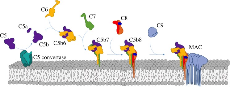
The membrane attack complex (MAC) is an important innate immune effector of the complement terminal pathway that forms cytotoxic pores on the surface of microbes. Despite many years of research, MAC structure and mechanism of action have remained elusive, relying heavily on modelling and inference from biochemical experiments. Recent advances in structural biology, specifically cryo-electron microscopy, have provided new insights into the molecular mechanism of MAC assembly. Its unique 'split-washer' shape, coupled with an irregular giant β-barrel architecture, enable an atypical mechanism of hole punching and represent a novel system for which to study pore formation. This review will introduce the complement terminal pathway that leads to formation of the MAC. Moreover, it will discuss how structures of the pore and component proteins underpin a mechanism for MAC function, modulation and inhibition.This article is part of the themed issue 'Membrane pores: from structure and assembly, to medicine and technology'.

Keywords: MACPF; cholesterol-dependent cytolysin; complement pathway; membrane attack complex; pore-forming protein; pore-forming toxins.


Conflict of interest statement

We declare we have no competing interests.

Figures

Figure 1.
Figure 1.
Illustration of the stepwise MAC assembly pathway from soluble complement factors. The first step requires cleavage of C5 (purple) into the small anaphylatoxin C5a and the large fragment C5b by the C5 convertase (turquoise). C6 (yellow) binds the labile C5b intermediate, resulting in a stable C5b6 complex. C7 (green) binds C5b6, anchoring the newly formed C5b7 complex to the membrane surface. C8, a heterotrimeric protein composed of C8α (orange), C8β (red) and C8γ (dark blue), is incorporated into the assembly precursor forming C5b8 and marking the first membrane penetrating event. Finally, multiple copies of C9 (light blue) join the assembly and span membrane, resulting in the final membrane attack complex (MAC).
Figure 2.
Figure 2.
Domain architecture of complement MACPF/CDC-containing proteins; C6, C7, C8α–γ, C8β, C9. (a) Schematic showing domain organization. MACPF domain is coloured in a combination of blue, red, green and yellow, consistent with the colouring used in the structures shown in (be). Regions that form the final β-barrel pore are indicated as TMH1 and TMH2. The ancillary domains are as follows: thrombospondin (TSP) (magenta), low-density lipoprotein receptor type A (LDLRA) (light pink), lipocalin (LIP) (dark blue), epidermal growth factor type (EGF) (black), complement control protein (CCP) (teal), and factor I-like module (FIM) (purple). (be) Crystal structures of soluble MAC components. (b) C6 (PDB ID: 3T5O). (c) C5b6 (PDB ID: 4A5W), where C6 is coloured as in (b) and C5b is shown in grey. (d) C8αγ component of the C8 heterotrimer (PDB ID: 3OJY). (e) C8β component of the C8 heterotrimer (PDB ID: 3OJY). (f) The two TMH regions are shown as clusters of α-helices in the soluble monomer protein. Upon a dramatic conformational change, the TMH regions unfurl into β-sheets that span the target membrane. Colours are as for sections (ae).
Figure 3.
Figure 3.
Comparable side and top views of polyC9, MAC and SC5b9 complexes determined using cryoelectron microscopy (cryo-EM). Pseudo-atomic model of a 22-fold symmetric C9 oligomer (PDB: 5FMW) (a) based on the polyC9 reconstruction (b) (EMBD: 3235). Alternating C9 monomers are coloured red and blue. Putative transmembrane regions (TM) are indicated by a black bar. (c) Cryo-EM reconstruction of the MAC pore (EMBD: 3134), where density for each protein component is coloured (see key). (d) Subtomogram average of the MAC on liposomes. Protein density is grey and lipid bilayer is orange (EMBD: 3289). (e) Soluble, regulated form of the MAC, SC5b9 (EMBD: 1991), in which non-MAC density is indicated by chaperones. Scale bar, 100 Å, is relevant for all structures.

Similar articles

Cited by

References

    1. Walport MJ. 2001. Complement. First of two parts. N. Engl. J. Med. 344, 1058–1066. (10.1056/NEJM200104053441406) - DOI - PubMed
    1. Bloch EF, et al. 1997. C5b-7 and C5b-8 precursors of the membrane attack complex (C5b-9) are effective killers of E. coli J5 during serum incubation. Immunol. Invest. 26, 409–419. (10.3109/08820139709022698) - DOI - PubMed
    1. Tomlinson S, Taylor PW, Morgan BP, Luzio JP. 1989. Killing of Gram-negative bacteria by complement. Fractionation of cell membranes after complement C5b-9 deposition on to the surface of Salmonella minnesota Re595. Biochem. J. 263, 505–511. (10.1042/bj2630505) - DOI - PMC - PubMed
    1. Harriman GR, Esser AF, Podack ER, Wunderlich AC, Braude AI, Lint TF, Curd JG. 1981. The role of C9 in complement-mediated killing of Neisseria. J. Immunol. 127, 2386–2390. - PubMed
    1. Ram S, Lewis LA, Rice PA. 2010. Infections of people with complement deficiencies and patients who have undergone splenectomy. Clin. Microbiol. Rev. 23, 740–780. (10.1128/CMR.00048-09) - DOI - PMC - PubMed

MeSH terms

Substances