. 2017 Oct;58(10):1988-1998.
doi: 10.1194/jlr.M077610. Epub 2017 Jun 19.

Cholesterol auxotrophy and intolerance to ezetimibe in mice with SREBP-2 deficiency in the intestine

Affiliations

Cholesterol auxotrophy and intolerance to ezetimibe in mice with SREBP-2 deficiency in the intestine

Shunxing Rong et al. J Lipid Res. 2017 Oct.

Abstract

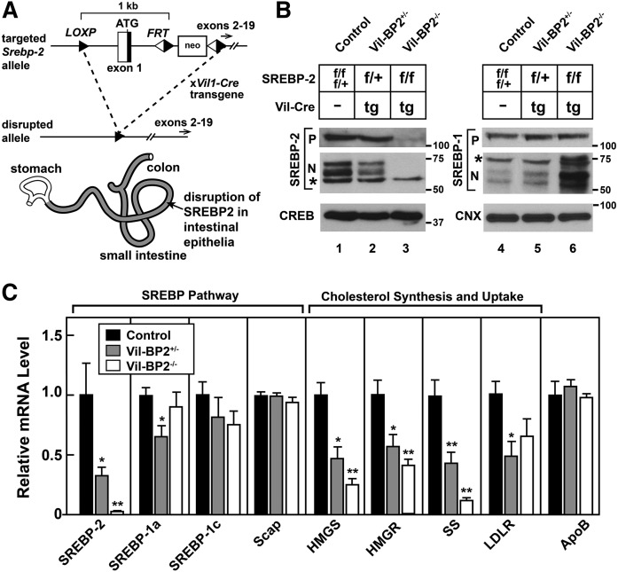
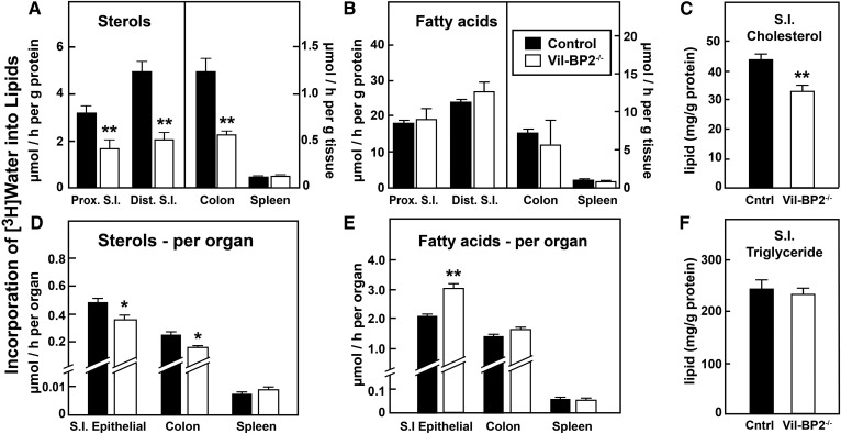
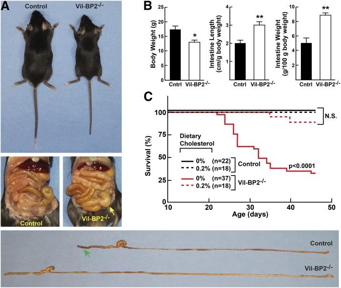
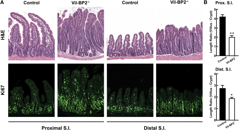
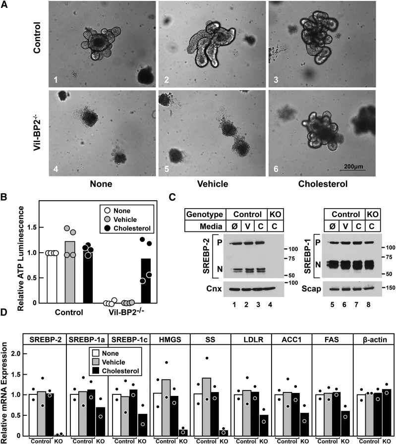
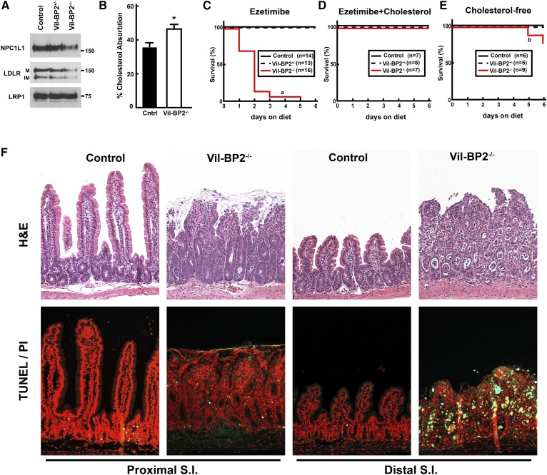
SREBP-2 activates transcription of all genes needed for cholesterol biosynthesis. To study SREBP-2 function in the intestine, we generated a mouse model (Vil-BP2-/- ) in which Cre recombinase ablates SREBP-2 in intestinal epithelia. Intestines of Vil-BP2-/- mice had reduced expression of genes required for sterol synthesis, in vivo sterol synthesis rates, and epithelial cholesterol contents. On a cholesterol-free diet, the mice displayed chronic enteropathy with histological abnormalities of both villi and crypts, growth restriction, and reduced survival that was prevented by supplementation of cholesterol in the diet. Likewise, SREBP-2-deficient enteroids required exogenous cholesterol for growth. Blockade of luminal cholesterol uptake into enterocytes with ezetimibe precipitated acutely lethal intestinal damage in Vil-BP2-/- mice, highlighting the critical interplay in the small intestine of sterol absorption via NPC1L1 and sterol synthesis via SREBP-2 in sustaining the intestinal mucosa. These data show that the small intestine requires SREBP-2 to drive cholesterol synthesis that sustains the intestinal epithelia when uptake of cholesterol from the gut lumen is not available, and provide a unique example of cholesterol auxotrophy expressed in an intact, adult mammal.

Keywords: Niemann-Pick C1-like 1; SREBP; Scap; cholesterol/biosynthesis; fatty acid/synthesis; organoid.


Figures

Fig. 1.
Fig. 1.
Disruption of Srebp-2 in intestinal epithelia. A: Schematic of conditional knockout of SREBP-2 in intestinal mucosa. Grayed areas indicate the zone of SREBP-2 disruption. B: Immunoblot analysis. Isolated intestinal epithelial cells (IECs) were prepared from mice of the indicated genotypes (female, 4–8 weeks of age, four or five mice per group). IECs were fractionated and equal amounts of protein from each mouse were pooled; 20 µg aliquots of the pooled membranes and nuclear extract were subjected to SDS-PAGE and immunoblot analysis. The precursor and nuclear forms of SREBPs are denoted as P and N, respectively. Asterisks denote nonspecific bands. Immunoblots of cAMP response element-binding protein (CREB) and calnexin (CNX) were used as loading controls for the nuclear extract and membrane fractions, respectively. Molecular mass in kDa is indicated. Mouse genotype is indicated, f, flox; +, wild-type; tg, transgenic. C: Relative mRNA levels. Total RNA from IECs from the same mice as used in B was isolated and subjected to QPCR. Each value represents the mean ± SEM of data from four or five mice. Values in littermates lacking Vil-Cre (the same mice used in lanes 1 and 4 of B, hereafter referred to as control mice) were arbitrarily defined as 1.0. Statistical significance between control and knockout mice in C was assessed by the two-tailed Student’s t-test, *P < 0.05, **P < 0.01.
Fig. 2.
Fig. 2.
Decreased sterol synthesis in SREBP-2-deficient intestine. A, B, D, E: Control and Vil-BP2−/− mice (male, 11–12 weeks of age, seven per group) were injected intraperitoneally with 3H-labeled water (50 mCi in 0.2 ml of saline). One hour later, tissues were removed and the small intestine was divided into two segments of equal length, proximal (Prox. S.I.) and distal (Dist. S.I.) from which IECs were immediately prepared. IECs or whole tissues (for spleen and colon) were then processed for isolation of digitonin-precipitable sterols (A, D) and FAs (B, E). The sterol and fatty acid synthetic rates were calculated as micromoles of 3H-radioactivity incorporated per hour per gram protein (small intestine IECs) or gram tissue (A, B) or per whole organ (D, E). C, F: Lipid contents in IECs of control and Vil-BP2−/− mice (male, 5 weeks of age, five per group). Each bar represents mean ± SEM from groups of mice. *P < 0.05, **P < 0.01 denotes the level of statistical significance (two-tailed Student’s t-test) between control and Vil-BP2−/− mice.
Fig. 3.
Fig. 3.
Disruption of intestinal SREBP-2 produces intestinal hypertrophy, enteropathy, and cholesterol auxotrophy. A: Gross appearance (top), abdominal organs (middle), and dissected gastrointestinal tracts (bottom) from control mice and Vil-BP2−/− mice are shown. Yellow arrow indicates protuberance of organs in Vil-BP2−/− mice. Green arrow indicates formed stools in colons of control mice absent in Vil-BP2−/− mice. B: Intestine length and weight (mice shown are the same as in Fig. 1B, C). C: Mice of the indicated genotypes (n per group is indicated in the key) and their dams were fed either a cholesterol-free chow diet or a diet containing 0.2% cholesterol from birth to 7 weeks of age. Survival percentage from age 10 days is shown. Statistical significance in B between groups of mice was assessed by the two-tailed Student’s t-test, *P < 0.05, and in C by Log-rank test (P value as indicated compares control and Vil-BP2−/− mice on cholesterol-free diet).
Fig. 4.
Fig. 4.
Crypt hyperplasia and villus atrophy of small intestinal mucosa in Vil-BP2−/− mice. A: Representative histologic sections of proximal (Proximal S.I.) and distal (Distal S.I.) small intestine from 4- to 5-week-old male control and Vil-BP2−/− mice stained with H&E or immunostained for Ki67; magnification, 100×. B: Length ratio of villus to crypt of proximal (Prox. S.I.) and distal (Dist. S.I.) small intestine. Lengths of five villi and crypts on an H&E-stained section were measured and averaged for each mouse. Length ratios from groups of control and Vil-BP2−/− mice (four mice per genotype) are shown. Each bar represents mean ± SEM from groups of mice. *P < 0.05, **P < 0.01 denotes the level of statistical significance (two-tailed Student’s t-test) between control and Vil-BP2−/− mice.
Fig. 5.
Fig. 5.
Cholesterol rescue of defective crypt growth in SREBP-2-deficient enteroids. Small intestinal crypts from control and Vil-BP2−/− mice were isolated, embedded in matrigel, and overlaid with media. Some wells received MβCD-cholesterol within the matrigel and in the media or equal concentrations of uncomplexed MβCD. Crypts receiving no additions (none, Ø), MβCD (vehicle, V), or MβCD-cholesterol (cholesterol, C) were then grown for 6 days. A: Representative enteroids were imaged under bright field microscopy (bar = 200 μm; magnification, 100×). B: Cell viability as measured by ATP luminescence (n = 4 per genotype). C: Immunoblot analysis. Whole-cell extracts prepared from enteroids (n = 2 per genotype) and equal amounts of protein from each mouse were pooled. Thirty microgram aliquots of pooled protein were subjected to SDS-PAGE and immunoblot analysis. The precursor and nuclear forms of SREBPs are denoted as P and N, respectively. Immunoblot of Cnx was used as a loading control. Molecular mass in kDa is indicated. D: Relative mRNA levels. Total RNA was isolated from enteroids and subjected to QPCR using cyclophilin as the invariant control. ACC1, acetyl-CoA carboxylase 1. For B and D, circles indicate the value of enteroids from each individual mouse per media condition and bars indicates the mean value. Data from Vil-BP2−/− enteroids that did not receive cholesterol are not shown in B and C as insufficient protein and RNA were recovered owing to enteroid death. The mean value of control enteroids without media additions is arbitrarily defined as 1.0 in B and D.
Fig. 6.
Fig. 6.
Mucosal injury after blockade of cholesterol absorption with ezetimibe in SREBP-2-deficient intestines. A: Immunoblot analysis. Pooled membrane fractions (20 µg per lane) from IECs (the same mice as those used in Fig. 1B, C) were subjected to SDS-PAGE and immunoblot analysis. M, mature form, IM, immature form of the LDLR, respectively. Immunoblot of LDL receptor-related protein 1 (LRP1) is used as a loading control. Molecular mass in kDa is indicated. B: Fractional cholesterol absorption. Control and Vil-BP2−/− mice (male, 5 weeks of age, five per group) were dosed orogastrically with a mixture of radiolabeled 3H-sitostanol and 14C-cholesterol. The 14C to 3H ratio in feces was then measured and used to calculate fractional cholesterol absorption; bars represent the mean ± SEM of data from five mice. C–E: Mice of the indicated genotypes (n per group as indicated in the key) were fed a cholesterol-free chow diet containing 0.01% ezetimibe either without (C) or with (D) 2% cholesterol and survival over the next 6 days was measured. A third group was fed the cholesterol-free chow without additions (E). F: Representative H&E- and TUNEL-stained; propidium iodide -costained histologic sections from proximal (Proximal S.I.) and distal (Proximal S.I.) small intestine from control and Vil-BP2−/− mice treated with ezetimibe for 2 days (magnification 100×). Statistical significance in B between groups of mice was assessed by the two-tailed Student’s t-test, *P < 0.05, and in C–E by Log-rank test (a compares control and Vil-BP2−/− mice fed ezetimibe, P < 0.01. b compares ezetimibe-fed and cholesterol-free diet fed Vil-BP2−/− mice, P < 0.01).

Comment in

Similar articles

Cited by

References

    1. Horton J. D., Goldstein J. L., and Brown M. S.. 2002. SREBPs: activators of the complete program of cholesterol and fatty acid synthesis in the liver. J. Clin. Invest. 109: 1125–1131. - PMC - PubMed
    1. Horton J. D., Shimomura I., Brown M. S., Hammer R. E., Goldstein J. L., and Shimano H.. 1998. Activation of cholesterol synthesis in preference to fatty acid synthesis in liver and adipose tissue of transgenic mice overproducing sterol regulatory element-binding protein-2. J. Clin. Invest. 101: 2331–2339. - PMC - PubMed
    1. Yang T., Espenshade P. J., Wright M. E., Yabe D., Gong Y., Aebersold R., Goldstein J. L., and Brown M. S.. 2002. Crucial step in cholesterol homeostasis: sterols promote binding of SCAP to INSIG-1, a membrane protein that facilitates retention of SREBPs in ER. Cell. 110: 489–500. - PubMed
    1. Ye J., and DeBose-Boyd R. A.. 2011. Regulation of cholesterol and fatty acid synthesis. Cold Spring Harb. Perspect. Biol. 3: a004754. - PMC - PubMed
    1. Rong S., Cortes V. A., Rashid S., Anderson N. N., McDonald J. G., Liang G., Moon Y. A., Hammer R. E., and Horton J. D.. 2017. Expression of SREBP-1c requires SREBP-2-mediated generation of a sterol ligand for LXR in livers of mice. eLife. 6: e25015. - PMC - PubMed

Publication types

MeSH terms

LinkOut - more resources