. 2017 Sep;92(3):318-326.
doi: 10.1124/mol.117.108894. Epub 2017 Jun 19.

Determination of the Residues in the Extracellular Domain of the Nicotinic α Subunit Required for the Actions of Physostigmine on Neuronal Nicotinic Receptors

Affiliations

Determination of the Residues in the Extracellular Domain of the Nicotinic α Subunit Required for the Actions of Physostigmine on Neuronal Nicotinic Receptors

Xiaochun Jin et al. Mol Pharmacol. 2017 Sep.

Abstract

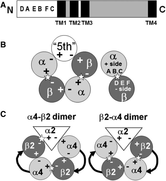
Physostigmine can potentiate and inhibit neuronal nicotinic receptors, in addition to inhibiting the activity of acetylcholinesterase. We found that receptors containing three copies of the α2 subunit are inhibited by low concentrations of physostigmine in contrast to receptors containing three copies of the α4 subunit that are potentiated. We exploited this observation to determine the regions required for the actions of physostigmine. Chimeric constructs of the α2 and α4 subunits located two regions in the extracellular amino-terminal domain of the subunit: the E loop (a loop of the transmitter-binding domain) and a region closer to the amino-terminus that collectively could completely determine the different effects of physostigmine. Point mutations then identified a single residue, α2(I92) versus α4(R92), that, when combined with transfer of the E loop, could convert the inhibition seen with α2 subunits to potentiation and the potentiation seen with α4 subunits to inhibition. In addition, other point mutations could affect the extent of potentiation or inhibition, indicating that a more extensive set of interactions in the amino-terminal domain plays some role in the actions of physostigmine.


Figures

Fig. 1.
Fig. 1.
Summary of subunit structure and arrangement in the pentameric receptor. The figure shows cartoon representations of the structure of neuronal nicotinic receptors. (A) Main structural regions of the primary sequence for both α and β subunits. Each subunit has a similar overall structure (A, bottom) with a relatively large N-terminal extracellular domain that contains the loops of the ACh-binding site. This is followed by three TMs; the channel is lined by the TM2 domains from each subunit. There is then a variable-length intracellular domain followed by a fourth TM and a short C-terminal extracellular domain. (B) The arrangement of subunits in the pentameric receptor viewed from the extracellular side (left); a canonical ACh-binding site (indicated by a star) is formed at an interface between an α subunit (“+” surface) counterclockwise to a β subunit (“−” surface). The α subunit contributes the A, B, and C loops (right), whereas the β subunit contributes the D, E, and F loops. (C) The location of the α2 subunit when expressed with the α4-β2 and β2-α4 dimeric constructs. Note that the nature of the interfaces formed by the α2 subunit depends on the particular constructs used.
Fig. 2.
Fig. 2.
Effect of transplanting the D-A and E loop regions between the α4 and α2 subunits. Current traces are shown for responses when ACh was initially applied alone, and then the perfusion was switched to ACh plus physostigmine (application of ACh alone is indicated by the bottom bar above the trace, and the time of the application of both ACh and physostigmine is shown by the top bar). The constructs injected are shown above the trace (20:1 ratio for α/β subunits); the horizontal bar shows 20 second for all traces, whereas the vertical bar shows the current calibration for each trace. The ACh concentration was adjusted to result in a response of less than 20% of the maximal response for that oocyte. The physostigmine concentration was 10 µM. The ACh concentrations used were as follows: α4β4, 10 µM; α2β4, 20 µM; α4(D-A+E)β4, 3 µM; and α2(D-A+E)β4, 10 µM.
Fig. 3.
Fig. 3.
The extracellular domains of α2 and α4 subunits. Sequences of the human α4 (accession number NP_000735.1) and mouse α2 (accession number NP_659052.1) subunits are shown for the amino-terminal extracellular region. In the line below the sequences (“Differ”) a “+” indicates that the residues differ at that position in the two subunits, whereas the approximate locations of the loops in the canonical ACh-binding site are shown above the sequences (“Loops”). Loops A, B, and C are contributed by the subunit at the “+” side of the interface, whereas loops D, E and F are contributed by the subunit on the “-“ side. The regions transferred in the various chimeras are indicated by the lines above the loops, and identified at the left (e.g., “A-TM1” starts at the end of the A loop and extends to the start of the first TM). Finally, the residues mutated in the D-A region are indicated by solid arrows below the sequences in the line marked “Mutations,” and the residues in the E loop are indicated by hollow arrows.
Fig. 4.
Fig. 4.
Effects of N-terminal region chimeras on the actions of physostigmine. The left-hand panel shows a cartoon representation of the structure of the N-terminal region of the various chimeras tested. Regions derived from the α2 subunit are shown in red, whereas regions from the α4 subunit are shown in blue. Regions that are identical between the two subunits are in gray. The chimeras are identified in the left-hand boxes; those based on the α2 subunit are shown in the bottom portion, whereas those based on the α4 subunit are shown in the top portion. The chimera joining points are shown in Fig. 3. The mean ± S.E. physostigmine effect is shown in the bar to the right, whereas the asterisks give the P value indicating that the effect is identical to the effect for the wild-type subunit (one-way ANOVA with Dunnett’s correction; ***P < 0.001, **P < 0.01). The solid vertical line shows an effect ratio of 1 (no effect), whereas the dashed lines show the mean effects for wild-type α2 and α4 subunits. Data values are given in Tables 3 and 5. All α subunits were expressed with β4 at a 20:1 ratio.
Fig. 5.
Fig. 5.
Effects of mutations in the D-A region. The figure is similar to Figure 4. In the left panel, a cartoon of the region from the D loop to the end of the E loop is shown, with regions derived from the α2 subunit in red and regions from the α4 subunit in blue. The A loop (gray) is identical in these subunits, whereas the region between the A and E loops (white) was not mutated. The mutations were made on the background of the α2(E) chimera (top four lines, hatched bars) or the α2(D-A+E) chimera (bottom 4 lines, solid black bars), whereas the middle four lines show results for relevant chimeras and wild-type α2 subunits. The approximate location of the residues mutated is shown by the colored bars shown in the cartoons (left). On the background of the α2(E) chimera, the mutation changed a residue found in the α2 sequence to that found in the α4 subunit and so is shown as red and vice versa. The dashed lines show the mean effects for the background chimeras, and the asterisks indicate the P value that the effect is identical to that for the background chimera (one-way ANOVA with Dunnett’s correction; ***P < 0.001, **P < 0.01). For locations of chimera joining points and mutations, see Fig. 3. Data are shown as the mean ± S.E.; data values are given in Table 4.
Fig. 6.
Fig. 6.
Location of mutated residues in the N-terminal domain of the α4 subunit. The figure shows views of the extracellular N-terminal domain of the nicotinic α4β2 receptors (Morales-Perez et al., 2016) to indicate the position of the residues mutated. The color coding is kept constant for all panels. (A) and (B) provide overall orientation. (A) A view from the extracellular fluid looking down at the receptor. The α4 subunits are shown as blue ribbons, whereas the β2 subunits are shown in green. The yellow arrows show the locations of canonical ACh-binding sites (α4 counterclockwise, contributing the “+” side). The red bracket indicates the β2-α4 interface shown in the remaining panels. (B) A side view of a β2-α4 pair from the outside of the receptor with the extracellular fluid at the top. The approximate position of the cell membrane is shown by the black bracket on the right. The E loop of the α4 subunit is shown in orange, and the D-A region is shown in cyan. (C) The region indicated by the red box in (B), enlarged and with the β2 subunit removed to reveal the α4 subunit. The view is from the outside of the receptor, looking into the cavity that binds drugs. The face of the “−” side of the binding pocket is shown. The peptide chain of the α4 subunit is shown as a blue ribbon, with the E loop shown in orange and the D-A region shown in cyan. The mutated residues are shown in stick format: V61 and H66 residues are shown in green to the right of the E loop; D76, Y77, and E78 residues are shown in black at the top of the panel; and R92 residue is shown in red behind the E loop. (D–F) Enlarged views of mutated residues. (D) The V61 and H66 residues (green stick format) seen from a perspective similar to (A). Although these residues are close to the E loop, they do not appear to be in a position to directly interact or extend into the drug-binding pocket. (E) The D76, Y77, and E78 residues (black stick format) seen from a perspective further into the extracellular milieu. These residues appear to be close to the initial α helix of the subunit and far from the E loop. (F) The R92 residue is shown in red seen from a perspective approximately 180° from that in (A), where R92 is behind the E loop and appears to approach the W123 sidechain in the E loop (shown in orange stick format).

References

    1. Hamouda AK, Kimm T, Cohen JB. (2013) Physostigmine and galanthamine bind in the presence of agonist at the canonical and noncanonical subunit interfaces of a nicotinic acetylcholine receptor. J Neurosci 33:485–494. - PMC - PubMed
    1. Harpsøe K, Ahring PK, Christensen JK, Jensen ML, Peters D, Balle T. (2011) Unraveling the high- and low-sensitivity agonist responses of nicotinic acetylcholine receptors. J Neurosci 31:10759–10766. - PMC - PubMed
    1. Jin X, Steinbach JH. (2011) A portable site: a binding element for 17β-estradiol can be placed on any subunit of a nicotinic α4β2 receptor. J Neurosci 31:5045–5054. - PMC - PubMed
    1. Jin X, Steinbach JH. (2015) Potentiation of neuronal nicotinic receptors by 17β-estradiol: roles of the carboxy-terminal and the amino-terminal extracellular domains. PLoS One 10:e0144631. - PMC - PubMed
    1. Jin X, McCollum MM, Germann AL, Akk G, Steinbach JH. (2017) The E loop of the transmitter binding site is a key determinant of the modulatory effects of physostigmine on neuronal nicotinic α4β2 receptors. Mol Pharmacol 91:100–109. - PMC - PubMed

Publication types

LinkOut - more resources