. 2017 Jul 3;114(27):E5370-E5378.
doi: 10.1073/pnas.1621279114. Epub 2017 Jun 19.

TCTE1 is a conserved component of the dynein regulatory complex and is required for motility and metabolism in mouse spermatozoa

Affiliations

TCTE1 is a conserved component of the dynein regulatory complex and is required for motility and metabolism in mouse spermatozoa

Julio M Castaneda et al. Proc Natl Acad Sci U S A. .

Abstract

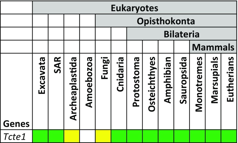
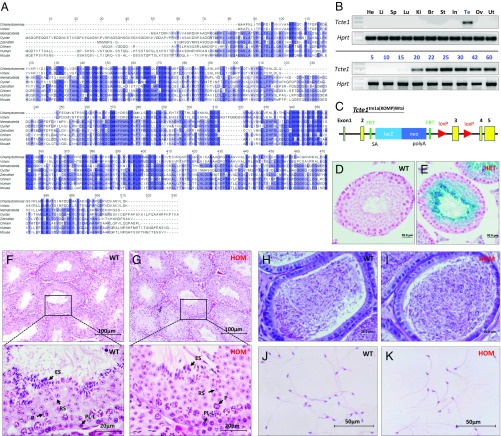
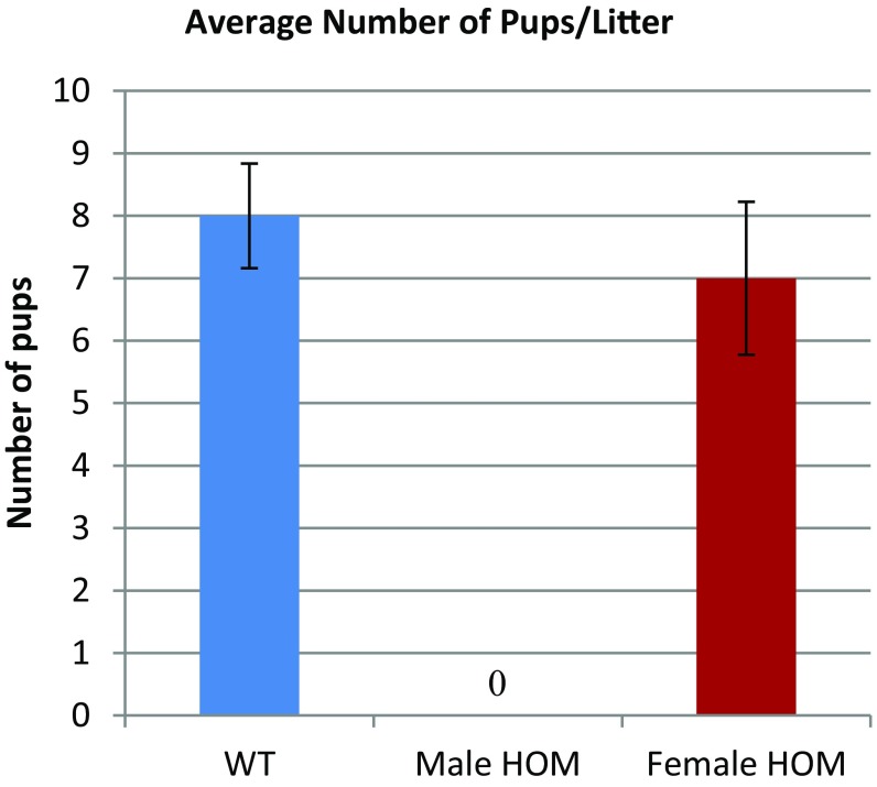
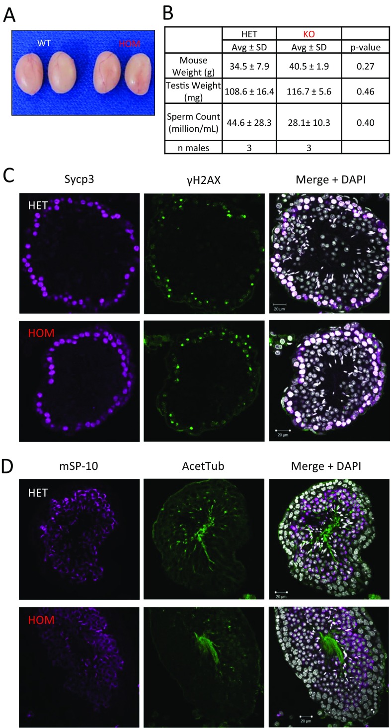
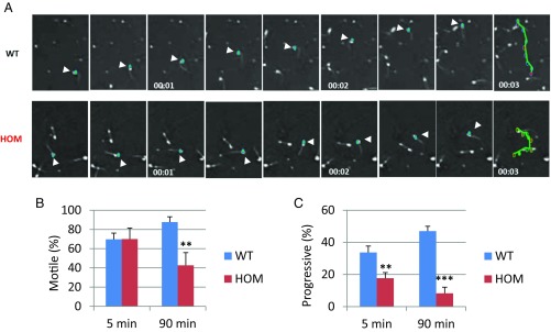
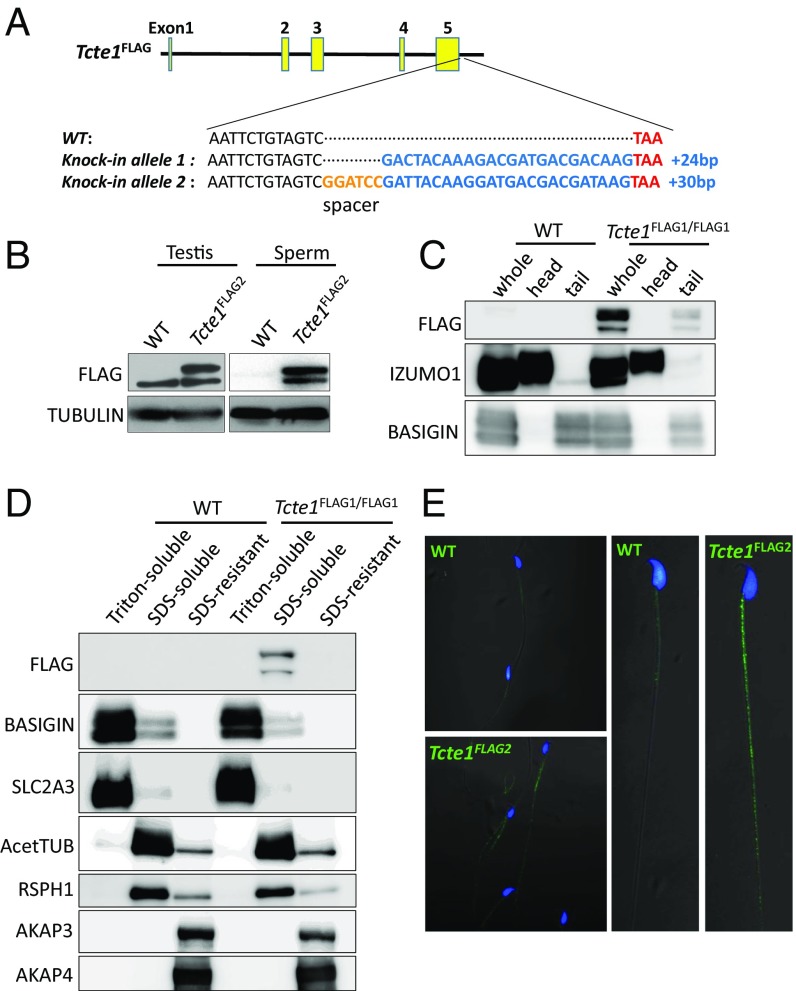
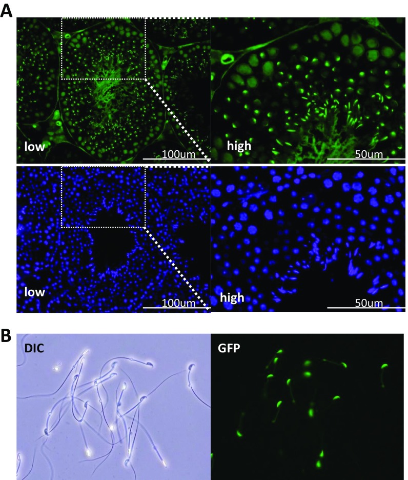
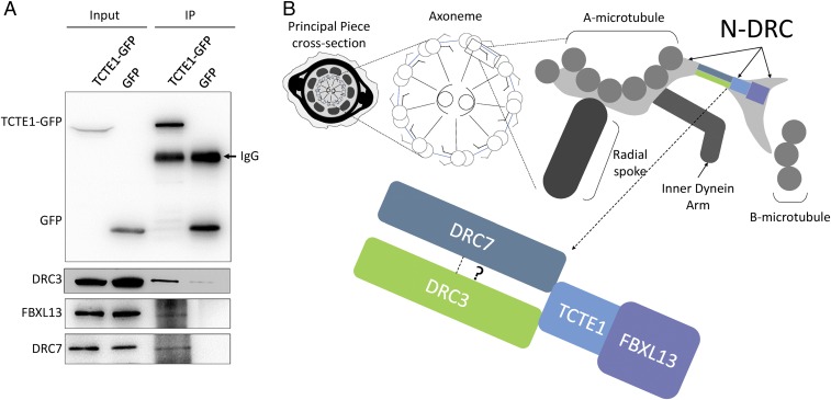
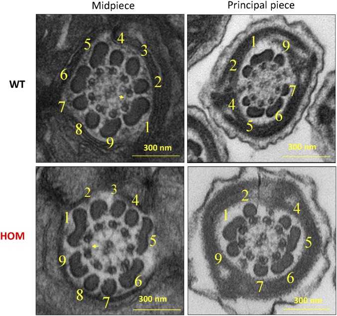
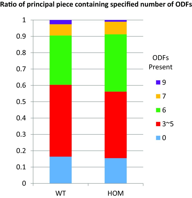
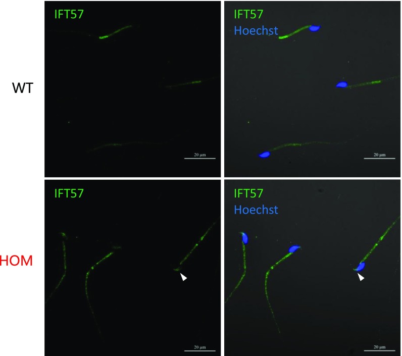
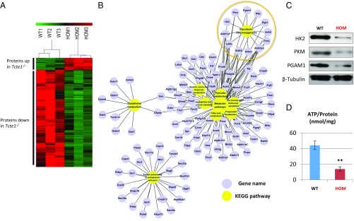
Flagella and cilia are critical cellular organelles that provide a means for cells to sense and progress through their environment. The central component of flagella and cilia is the axoneme, which comprises the "9+2" microtubule arrangement, dynein arms, radial spokes, and the nexin-dynein regulatory complex (N-DRC). Failure to properly assemble components of the axoneme leads to defective flagella and in humans leads to a collection of diseases referred to as ciliopathies. Ciliopathies can manifest as severe syndromic diseases that affect lung and kidney function, central nervous system development, bone formation, visceral organ organization, and reproduction. T-Complex-Associated-Testis-Expressed 1 (TCTE1) is an evolutionarily conserved axonemal protein present from Chlamydomonas (DRC5) to mammals that localizes to the N-DRC. Here, we show that mouse TCTE1 is testis-enriched in its expression, with its mRNA appearing in early round spermatids and protein localized to the flagellum. TCTE1 is 498 aa in length with a leucine rich repeat domain at the C terminus and is present in eukaryotes containing a flagellum. Knockout of Tcte1 results in male sterility because Tcte1-null spermatozoa show aberrant motility. Although the axoneme is structurally normal in Tcte1 mutant spermatozoa, Tcte1-null sperm demonstrate a significant decrease of ATP, which is used by dynein motors to generate the bending force of the flagellum. These data provide a link to defining the molecular intricacies required for axoneme function, sperm motility, and male fertility.

Keywords: asthenozoospermia; glycolysis; male infertility; mutant mouse; testis-specific gene.


Conflict of interest statement

The authors declare no conflict of interest.

Figures

Fig. S1.
Fig. S1.
Evolution of Tcte1 among eukaryotes. Tcte1 is present in most eukaryotes that contain flagella (SAR: stramenopiles, alveolates, Rhizaria). Green indicates presence of Tcte1 in most species within taxon. Yellow indicates loss in several species within taxon.
Fig. 1.
Fig. 1.
Tcte1 is a conserved testis-enriched gene required for male fertility. (A) Sequence alignment of TCTE1 proteins from several metazoans and two volvocaceaen species. Only the LRR domain is shown. (B, Top panels) RT-PCR for Tcte1 in various mouse tissues with Hprt as a control. (Bottom panels) RT-PCR from testis at various postnatal days with Hprt as a control. (C) Schematic of the Tcte1tm1a(KOMP)Wtsi (Tcte1) allele from Wellcome Trust Sanger Institute. (D and E) β-Galactosidase staining of wild-type (D) and Tcte1+/− testis (E). (F and G) H&E-stained testis cross-sections from control (F) and Tcte1−/− mice (G). (Bottom panels in F and G: magnification of Top panels in F and G) Arrows highlight germ cells at various stages of spermatogenesis. PL-L, preleptonema-leptonema; P, pachynema; RS, round spermatid; ES, elongated spermatid. (H and I) periodic acid-Schiff (PAS)-stained cauda epididymis sections from control (H) and Tcte1−/− mice (I). (J and K) Spermatozoa from control (J) and Tcte1−/− mice (K).
Fig. S2.
Fig. S2.
Tcte1 HOM mutant females are fertile. Bar graph showing the average number of pups/litter between wild-type males, Tcte1-null males, and Tcte1-null females (n = 5).
Fig. S3.
Fig. S3.
Spermatogenesis appears normal in Tcte1−/− mice. (A) Testes from 9-wk-old control and KO mice. (B) Average mouse weight, testis weight, and sperm counts from three control and three KO males. (C) Meiotic markers Sypc3 and γH2AX are comparable in both control and KO testis cross-sections. (D) The acrosome caps (mSP-10) and acet-Tubulin are comparable in both control and KO testis cross-sections.
Fig. 2.
Fig. 2.
Tcte1 null males display asthenozoospermia. (A) Movie frames from recordings of spermatozoa from a control and homozygous mutant male. Teal: sperm head; green: path of an individual spermatozoon (arrowheads highlight sperm head). (B) Percentage of motile sperm from control and homozygous males at 5 min and 90 min (**P < 0.01). (C) Percentage of progressively motile sperm from control and homozygous males at 5 min and 90 min (**P < 0.01, ***P < 0.001).
Fig. 3.
Fig. 3.
TCTE1 localizes to sperm tails. (A) Schematic of FLAG-tagged alleles of endogenous Tcte1 generated using CRISPR/Cas9. FLAG2 allele has a 2-aa (Gly-Ser) spacer. (B) Western blot analysis of protein extracts from testis and sperm from wild type and Tcte1FLAG2/FLAG2 mice. (C) Western blot analysis of spermatozoa fractionated into sperm heads and sperm tails from wild-type and Tcte1FLAG1/FLAG1 mice. (D) Western blot analysis of sperm fractionated into Triton X-100 soluble, SDS soluble, and SDS insoluble fractions from wild-type and Tcte1FLAG1/FLAG1 mice. (E) Immunofluorescence analysis of spermatozoa from wild-type and Tcte1FLAG2/FLAG2 mice using anti-FLAG antibody (green: anti-FLAG signal; Hoechst: blue).
Fig. S4.
Fig. S4.
Tcte1FLAG2/FLAG2 males are fertile. Breeding results from Tcte1FLAG2/+ and Tcte1FLAG2/FLAG2 males paired with wild-type females.
Fig. S5.
Fig. S5.
Tcte1FLAG1 spermatogenesis appears normal. Analysis of spermatogenesis in Tcte1FLAG1/FLAG1 males generated with ES cells expressing GFP ubiquitously and within the acrosome. (A) Cross-sections from seminiferous tubules from Tcte1FLAG1/FLAG1 chimeric males. Top panels show GFP expression ubiquitously and in the acrosome; Bottom panels show Hoechst staining. (B) Differential interference contrast and epifluorescent images of spermatozoa from Tcte1FLAG1/FLAG1 males.
Fig. 4.
Fig. 4.
TCTE1 is a component of the dynein regulatory complex. (A) Individual DRC components were coexpressed in HEK293T cells. Immunoprecipitation of TCTE1-GFP coimmunoprecipitates DRC3, -6, and -7 (Bottom panels, third lane). (B) Model of TCTE1 localization within the DRC (adapted from ref. 23).
Fig. 5.
Fig. 5.
Flagellum ultrastructure appears normal. Electron micrographs showing cross-sections of the midpiece (Left panels) and principal piece (Right panels) of sperm from wild-type and Tcte1−/− mice. Numbers highlight the outer dense fibers, and yellow arrows highlight the presence of nexin links between microtubules.
Fig. S6.
Fig. S6.
The number of outer dense fibers in Tcte1−/− spermatozoa appears normal. EM cross-sections of the principal piece of sperm were quantified and categorized according to the number of outer dense fibers (ODFs) present [wild type (WT): n = 116, HOM: n = 194].
Fig. S7.
Fig. S7.
Aberrant intraflagellar transport localization. Immunofluorescence staining against IFT57 (a component of the intraflagellar transport machinery) shows persistent staining along the flagellum of Tcte1 HOM-null spermatozoa, including in the head (white arrowhead).
Fig. 6.
Fig. 6.
Glycolysis is decreased in Tcte1-null sperm. (A) Heat map from three independent proteomic analyses of sperm from wild-type and Tcte1-null mice. Red: proteins up-regulated; green: proteins down-regulated. (B) Grouping of proteins down-regulated in Tcte1-null sperm according to metabolic pathways. (C) Measured levels of ATP between wild-type and Tcte1-null sperm (n = 3). (D) Western blot analysis of three glycolytic enzymes from wild-type and Tcte1-null sperm.

References

    1. Blocker A, Komoriya K, Aizawa S. Type III secretion systems and bacterial flagella: Insights into their function from structural similarities. Proc Natl Acad Sci USA. 2003;100:3027–3030. - PMC - PubMed
    1. Carvalho-Santos Z, Azimzadeh J, Pereira-Leal JB, Bettencourt-Dias M. Evolution: Tracing the origins of centrioles, cilia, and flagella. J Cell Biol. 2011;194:165–175. - PMC - PubMed
    1. Faguy DM, Jarrell KF, Kuzio J, Kalmokoff ML. Molecular analysis of archael flagellins: Similarity to the type IV pilin-transport superfamily widespread in bacteria. Can J Microbiol. 1994;40:67–71. - PubMed
    1. Lindemann CB, Lesich KA. Functional anatomy of the mammalian sperm flagellum. Cytoskeleton. 2016;73:652–669. - PubMed
    1. Allen RD. A reinvestigation of cross-sections of cilia. J Cell Biol. 1968;37:825–831. - PMC - PubMed

Publication types

MeSH terms