. 2017 Aug 15;6(8):1137-1148.
doi: 10.1242/bio.022723.

Apical and basal epitheliomuscular F-actin dynamics during Hydra bud evagination

Affiliations

Apical and basal epitheliomuscular F-actin dynamics during Hydra bud evagination

Roland Aufschnaiter et al. Biol Open. .

Abstract

Bending of 2D cell sheets is a fundamental morphogenetic mechanism during animal development and reproduction. A critical player driving cell shape during tissue bending is the actin cytoskeleton. Much of our current knowledge about actin dynamics in whole organisms stems from studies of embryonic development in bilaterian model organisms. Here, we have analyzed actin-based processes during asexual bud evagination in the simple metazoan Hydra We created transgenic Hydra strains stably expressing the actin marker Lifeact-GFP in either ectodermal or endodermal epitheliomuscular cells. We then combined live imaging with conventional phalloidin staining to directly follow actin reorganization. Bending of the Hydra epithelial double layer is initiated by a group of epitheliomuscular cells in the endodermal layer. These cells shorten their apical-basal axis and arrange their basal muscle processes in a circular configuration. We propose that this rearrangement generates the initial forces to bend the endoderm towards the ectoderm. Convergent tissue movement in both epithelial layers towards the centre of evagination then leads to elongation and extension of the bud along its new body axis. Tissue movement into the bud is associated with lateral intercalation of epithelial cells, remodelling of apical septate junctions, and rearrangement of basal muscle processes. The work presented here extends the analysis of morphogenetic mechanisms beyond embryonic tissues of model bilaterians.

Keywords: Cnidarian; Epithelial cell; Evolution; Lifeact; Morphogenesis; Tissue evagination.


Conflict of interest statement

Competing interestsThe authors declare no competing or financial interests.

Figures
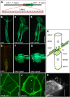
Fig. 1.
Fig. 1.
Transgenic Hydra strains expressing Lifeact-GFP. (A) Codon-optimized sequence of the lifeact-GFP expression construct driven by the Hydra actin1 promoter. (B) Hydra polyp with mosaic expression of Lifeact-GFP in large areas of the ectodermal layer. (C) Cluster of three transgenic ectodermal epithelial cells with focus level at the basal muscle processes running along the primary oral-aboral axis; (D) the same cell cluster with focus at apical cell junctions. (E) Hydra polyp with mosaic expression of Lifeact-GFP in the endodermal layer. (F) Cluster of four transgenic endodermal epithelial cells with focus level on basal muscle processes; (G) the same cell cluster with focus at apical cell junctions. (H) Schematic drawing of an ectodermal and an endodermal epithelial cell as positioned in the body wall of Hydra with prominent actin structures highlighted in green. Basal muscle processes in ectodermal and endodermal epithelial cells are oriented perpendicular to each other. The mesoglea linking both tissue layers is not included. The connections between ectodermal and endodermal epithelial cells across the mesoglea are also not shown. (I-K) F-actin structures at the apical surface of ectodermal epithelial cells. (I) Confocal projection (3.5 µm depth) at apical cell junctions in fixed tissue stained with Alexa Fluor 488 Phalloidin. (J) Corresponding confocal section at apical cell junctions in a living mosaic Lifeact-GFP polyp. (K) Actin-based apicolateral lamellipodia-like structures (arrows) as visualized in a single frame of a movie taken with a TIRF microscope in a living mosaic Lifeact-GFP polyp (see Movie 2). The black space around the transgenic cell is occupied by nontransgenic epithelial cells. Scale bars: 50 µm in C, D, F and G; 10 µm in I-K.
Fig. 2.
Fig. 2.
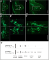
Early circular arrangement of endodermal muscle processes. (A,B,C) Confocal projections of the basal regions of ectodermal and endodermal epithelial cells of the budding zone stained with Alexa Fluor 488 Phalloidin. (A) Late stage 1 bud, in which ectodermal muscle processes run vertically and endodermal ones run horizontally. In the centre, endodermal muscle processes start to deviate from their regular orientation and appear shortened. (B) Eye-shaped arrangement of endodermal muscle processes in bud stage 1-2. (C) Circular orientation of endodermal muscle processes in bud stage 2, which reflects the correct endodermal planar polarity within the newly forming polyp. (A′,B′,C′) Orthogonal optical sections of (A,B,C) at the positions indicated by arrows show that endodermal bending into the ectodermal layer is correlated with circular arrangement of endodermal muscle processes. (D) Scheme of the arrangement of endodermal muscle processes at bud stage 2. ENML, endodermal muscle layer; M, mesoglea; EC, ectodermal layer. (E) Timing of endodermal muscle process reorientation according to bud stages from Otto and Campbell (1977). Depth of confocal projections: 20 µm in A; 15 µm in B; 25 µm in C. Scale bars: 50 µm in A and B; 20 µm in C.
Fig. 3.
Fig. 3.
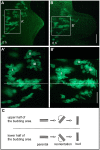
Lateral views of early bud stages showing ectodermal muscle process reorientation. (A,B) Confocal projections of specimens stained with Alexa Fluor 488 Phalloidin. (A′,B′) Schematic drawings of ectodermal muscle processes (dashed lines) at the corresponding stages. While ectodermal muscle processes moving into the bud from oral and aboral directions are oriented rather correctly with respect to the buds new body axes, those entering the bud from lateral areas have to increasingly rearrange in order to attain proper orientation. This results in a triangle shape at bud stage 3-4. (C) Timing of ectodermal muscle process reorientation according to bud stages from Otto and Campbell (1977). Projection depths: 30 µm in A; 10 µm in B. ECML, ectodermal muscle layer; ES, ectodermal apical surface. Scale bars: 50 µm.
Fig. 4.
Fig. 4.
Reorientation of ectodermal muscle processes during bud evagination. (A,B,C) Live imaging of a developing bud at three time points (0, 4, 8 h) showing muscle processes in patches of transgenic cells. (A′,B′,C′) Magnified images reveal the rotational direction and contraction during reorientation in the upper (oriented towards the head of the mother polyp) and lower (oriented towards the foot of the mother polyp) half of the bud. Stars and triangles label two individual muscle processes in order to demonstrate converging movement. (D) Schematic summary of the events. Scale bars: 200 µm in A, B and C; 100 µm in A′, B′ and C′.
Fig. 5.
Fig. 5.
Reorientation of endodermal muscle processes during bud evagination. (A,B) Live imaging of a developing bud at two time points (0, 6 h) showing muscle processes in patches of transgenic cells. (A′,B′) Magnified images reveal the rotational direction during reorientation in the upper (oriented towards the head of the mother polyp) and lower (oriented towards the foot of the mother polyp) half of the bud. (C) Schematic summary of the events. Scale bars: 200 µm in A and B; 100 µm in A′ and B′.
Fig. 6.
Fig. 6.
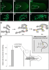
Polarized remodelling of ectodermal apical cell junctions during bud evagination. (A,B,C) Live imaging of a developing bud at three time points (0, 4, 8 h) showing a larger patch of transgenic cells. (A′,B′,C′) Magnified images are focused on F-actin associated with apical septate junctions; individual transgenic cells are labelled with numbers. (D) Schematic drawings of the transgenic cells in A′, B′ and C′, highlighting their positions and contact areas. Yellow dots mark new cell-cell contacts, which are established along the newly developing oral-aboral axis of the bud. Red circles mark detachment of existing cell-cell contacts, oriented perpendicular to the oral-aboral axis of the bud. (E) Relative rate of apical junctional remodelling in the evaginating area (grey bar) as compared with control tissue in the mid-gastric region (white bar). To determine the rate of remodelling, the number of polarized changes (yellow dots + red circles) per existing cell-cell contact between two transgenic ectodermal epithelial cells was counted during a period of 12 h. For evaginating cells, a total of six cell clusters including 110 cell-cell contacts were analyzed. During the tracking period, these cells moved from roughly −200 µm distance to the bud-parent border within the mother polyp to roughly 300 µm distance to the bud-parent border in the bud, and about one out of two cells either connected to a new cell neighbour or lost contact to its previous neighbour. For control cells, a total of three cell clusters including 157 cell-cell contacts were analyzed. These cells showed much slower movement along the oral-aboral axis of the mother polyp, and changed their neighbours at a very low rate. Scale bars: 200 µm in A, B and C; 100 µm in A′, B′ and C′.
Fig. 7.
Fig. 7.
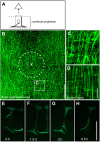
Randomly oriented ectodermal muscle processes in the larger budding area. (A) Schematic showing the perspective of the observer on the plane of the epithelium along an outside-inside direction. Confocal projection for B-D starts directly below the apical septate junctions of ectodermal epithelial cells and extends towards the basal end of endodermal epithelial cells including their muscle layer. Samples were stained with Alexa Fluor 488 Phalloidin. (B) Confocal projection of a stage 2-3 bud and the surrounding tissue showing a large number of muscle processes bent or randomly oriented. X marks the centre of evagination; the dotted circle indicates the bud-parent border. (C) Magnified view as indicated in B. (D) Regular arrangement of muscle processes in unevaginated control tissue from the mid-gastric region. (E-H) Short interval live tracking of an individual ectodermal epithelial cell during tissue recruitment shows dynamic remodelling of its actin fibers. Scale bars: 100 µm in B; 20 µm in C and D; 100 µm in E-H.
Fig. 8.
Fig. 8.
Actin-based processes involved in bud morphogenesis. (A) Bending. Circular arrangement and contractile activity of basal muscle processes in a group of endodermal epithelial cells may participate in initiating tissue bending. The blue line in the lower scheme represents the mesoglea, and the ectodermal layer is not shown. The confocal image on a stage 2 bud is taken from Philipp et al. (2009) with permission from PNAS. (B) Tissue recruitment. Movement of epithelial cells towards the centre of evagination during bud elongation and tissue recruitment involves apical junction remodelling and retraction, re-polarization and re-attachment onto the mesoglea of basal muscle processes.

References

    1. Anton-Erxleben F., Thomas A., Wittlieb J., Fraune S. and Bosch T. C. G. (2009). Plasticity of epithelial cell shape in response to upstream signals: a whole-organism study using transgenic Hydra. Zoology 112, 185-194. 10.1016/j.zool.2008.09.002 - DOI - PubMed
    1. Aufschnaiter R., Zamir E. A., Little C. D., Özbek S., Münder S., David C. N., Li L., Sarras M. P. Jr. and Zhang X. (2011). In vivo imaging of basement membrane movement: ECM patterning shapes Hydra polyps. J. Cell Sci. 124, 4027-4038. 10.1242/jcs.087239 - DOI - PMC - PubMed
    1. Burkel B. M., von Dassow G. and Bement W. M. (2007). Versatile fluorescent probes for actin filaments based on the actin-binding domain of utrophin. Cell Motil. Cytoskel. 64, 822-832. 10.1002/cm.20226 - DOI - PMC - PubMed
    1. Broun M., Gee L., Reinhardt B. and Bode H. R. (2005). Formation of the head organizer in hydra involves the canonical Wnt pathway. Development 132, 2907-2916. 10.1242/dev.01848 - DOI - PubMed
    1. Campbell R. D. (1967). Cell behavior and morphogenesis in hydroids. In Vitro 3, 22-32. 10.1007/BF02615918 - DOI

LinkOut - more resources