. 2017 Jun 19;7(1):3766.
doi: 10.1038/s41598-017-02756-3.

The role of Antibody Vκ Framework 3 region towards Antigen binding: Effects on recombinant production and Protein L binding

Affiliations

The role of Antibody Vκ Framework 3 region towards Antigen binding: Effects on recombinant production and Protein L binding

Chinh Tran-To Su et al. Sci Rep. .

Abstract

Antibody research has traditionally focused on heavy chains, often neglecting the important complementary role of light chains in antibody formation and secretion. In the light chain, the complementarity-determining region 3 (VL-CDR3) is specifically implicated in disease states. By modulating VL-CDR3 exposure on the scaffold through deletions in the framework region 3 (VL-FWR3), we further investigated the effects on secretion in recombinant production and antigen binding kinetics. Our random deletions of two residues in the VL-FWR3 of a Trastuzumab model showed that the single deletions could impact recombinant production without significant effect on Her2 binding. When both the selected residues were deleted, antibody secretion was additively decreased, and so was Her2 binding kinetics. Interestingly, we also found allosteric effects on the Protein L binding site at VL-FWR1 elicited by these deletions in VL- FWR3. Together, these findings demonstrate the importance of light chain FWR3 in antigen binding, recombinant production, and antibody purification using Protein L.


Conflict of interest statement

The authors declare that they have no competing interests.

Figures

Figure 1
Figure 1
The Trastuzumab mutants (WT, delT, delE, delTE) used in this study (A) Sequence alignments of the Trastuzumab mutants with CDRs highlighted as follows: Vκ-CDR1 (cyan), Vκ-CDR2 (magenta), and Vκ-CDR3 (orange). The deleted residues T74 and E81 are in blue and green, respectively. The structure of the WT-Fab region is shown in dark gray (heavy chain) and in light gray (κ light chain). Schematics of Her2 binding (at CDRs) and protein L binding (at Vκ-FWR1) are shown. (B) Antibody secretion levels of Trastuzumab mutants as determined by Protein G and L biosensor. Each bar shows the average concentration (μg/ml) of Trastuzumab WT and mutants in cell culture supernatants from three independent experiments using protein G and protein L biosensors, followed by statistical analysis using ANOVA and two tailed T-Test. The ANOVA and T-test results showed that the mutants are significantly different from one another (p < 0.05) within each biosensor data set (i.e. Protein L or G).
Figure 2
Figure 2
Binding kinetics profiles of Trastuzumab WT and its mutants from three independent experiments with corresponding average association (Ka), dissociation (Kd) and overall binding kinetic (KD) values with standard deviations. (A) Binding kinetics of decreasing concentration (100 nM to 6.25 nM) of Trastuzumab mutants to protein L biosensor, and (B) binding kinetics of Trastuzumab mutants using anti-human Fc capture biosensor to varying concentrations of Her2 (100 nM to 6.25 nM). Poor binding responses of the delTE mutant against protein L and Her2 produced unreliable binding kinetics values as determined by the Octet software for response value < 0.1.
Figure 3
Figure 3
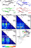
Results of the structural analyses of the Trastuzumab mutants FWRs and CDRs. (A) Damages in the Vκ-FWR1 β-strands (residues 5–22) subsequently caused conformational changes in the interacting regions and diminished the binding of protein L (Figs S3 and S4). (B) Contact maps of the Fab mutant-Her2 complexes against the Fab WT-Her2 complex showed common interactions (black dots) and WT-only interactions (magenta dots). Vκ-CDR1 and Vκ-CDR3 contacts with Her2 are shown in the boxes. Conformational changes of the Vκ-CDR3 loop with different orientations of Y92WT are shown (exposure is shown in Fig. S5B of Supplementary Materials). The mutants are colored as in (A): WT (black), delT (blue), delE (green) and delTE (red). All models were made using I-TASSER with PDB: 1n8z as template and minimized using AMBER14.

References

    1. Vidarsson, G., Dekkers, G. & Rispens, T. IgG subclasses and allotypes: from structure to effector functions. Front. Immunol. 5, doi:10.3389/fimmu.2014.00520 (2014). - PMC - PubMed
    1. Brochet X, Lefranc M, Giudicelli V. IMGT/V-QUEST: the highly customized and integrated system for IG and TR standardized V-J and V-D-J sequence analysis. Nucleic Acids Res. 2008;36:W503–508. doi: 10.1093/nar/gkn316. - DOI - PMC - PubMed
    1. Giudicelli V, Brochet X, Lefranc M. IMGT/V-QUEST: IMGT standardized analysis of the immunoglobulin (IG) and T cell receptor (TR) nucleotide sequences. Cold Spring Harb Protoc. 2011;2011:695–715. - PubMed
    1. Elgert, K. D. In Immunology - Understanding the Immune System (John Wiley & Sons, Inc., 1998).
    1. Jemmerson R, Mueller C, Flaa D. Differences in heavy chain amino acid sequences affecting the specificity of antibodies for variants of cytochrome c. Mol Immunol. 1993;30:1107–1114. doi: 10.1016/0161-5890(93)90157-7. - DOI - PubMed

Publication types

MeSH terms