. 2017:2017:8932583.
doi: 10.1155/2017/8932583. Epub 2017 May 28.

An Integrative Developmental Genomics and Systems Biology Approach to Identify an In Vivo Sox Trio-Mediated Gene Regulatory Network in Murine Embryos

Affiliations

An Integrative Developmental Genomics and Systems Biology Approach to Identify an In Vivo Sox Trio-Mediated Gene Regulatory Network in Murine Embryos

Wenqing Jean Lee et al. Biomed Res Int. 2017.

Abstract

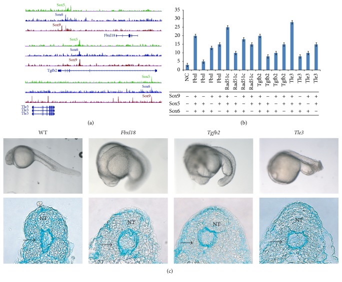
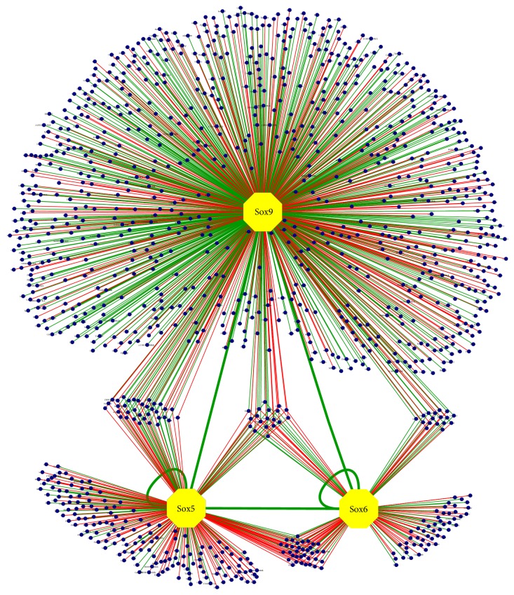
Embryogenesis is an intricate process involving multiple genes and pathways. Some of the key transcription factors controlling specific cell types are the Sox trio, namely, Sox5, Sox6, and Sox9, which play crucial roles in organogenesis working in a concerted manner. Much however still needs to be learned about their combinatorial roles during this process. A developmental genomics and systems biology approach offers to complement the reductionist methodology of current developmental biology and provide a more comprehensive and integrated view of the interrelationships of complex regulatory networks that occur during organogenesis. By combining cell type-specific transcriptome analysis and in vivo ChIP-Seq of the Sox trio using mouse embryos, we provide evidence for the direct control of Sox5 and Sox6 by the transcriptional trio in the murine model and by Morpholino knockdown in zebrafish and demonstrate the novel role of Tgfb2, Fbxl18, and Tle3 in formation of Sox5, Sox6, and Sox9 dependent tissues. Concurrently, a complete embryonic gene regulatory network has been generated, identifying a wide repertoire of genes involved and controlled by the Sox trio in the intricate process of normal embryogenesis.


Figures

Figure 1
Figure 1
Sox9, Sox5, and Sox6 gene targeting and their E13.5 transgenic embryos generated for FACS and expression Profiling. (a) Wildtype Sox9 allele (Sox9+) is indicated with exons depicted as black boxes. Translation start site (ATG) as indicated. (b) Sox9 with the F2A-EGFP-FRTNeo inserted in exon 3 (Sox9+(EGFPNeo)) and after FLPe recombination (Sox9+(EGFP)). Sox9+/+(EGFP) E13.5 embryo shown on the right. (c) Sox9-null allele with the EGFP-loxPNeo inserted after the ATG (Sox9−(EGFPNeo)) and after CRE recombination (Sox9−(EGFP)). Sox9-null allele with FRTNeo inserted in exon 1 (Sox9−(Neo)) and after FLPe recombination (Sox9). Sox9−/−(EGFP) E13.5 chimeric embryo shown on the right. (d) Exon containing the ATG of wildtype Sox5 (Sox5+) shown. EGFP-loxPNeo inserted after the ATG (Sox5−(EGFPNeo)) and after CRE recombination (Sox5−(EGFP)). Sox5+/−(EGFP) E13.5 embryo shown on the right. (e) Exon containing the ATG of wildtype Sox6 (Sox6+) shown. EGFP-loxPNeo inserted after the ATG (Sox6−(EGFPNeo)) and after CRE recombination (Sox6−(EGFP)). Sox6+/−(EGFP) E13.5 embryo shown with a wildtype embryo (WT) under same lighting condition. EGFP, green boxes; F2A, blue ovals; FRT sites, red triangles; loxP sites, yellow triangles (see also Figure S1 and Table S1).
Figure 2
Figure 2
Differentially expressed genes from the Sox9 microarray and Sox5/Sox6 double-null microarray. (a) Top 10 GO-terms for genes that are downregulated when Sox9 is inactivated. (b) Top 10 GO-terms for genes that are downregulated when Sox5 and Sox6 are inactivated (see also Figure S2 and Table S2).
Figure 3
Figure 3
ChIP-seq results for Sox9, Sox5, and Sox6. (a) Dissected tissues of the E13.5 embryo used for ChIP are indicated by the dotted blue lines. (b) Distribution of the Sox9 binding sites with respect to their nearest genes. (c) Primary Sox9 motif found shows Sox9 acts as a homodimer with the binding sites in opposite orientation. (d) Primary motif found from the Sox5 binding profile. (e) Primary motif found from the Sox6 binding profile. (f) Distribution of the Sox5 binding sites with respect to their nearest genes. (g) Distribution of the Sox6 binding sites with respect to their nearest genes (see also Figure S3 and Table S3).
Figure 4
Figure 4
(a) Top 10 GO-terms for genes that are activated by Sox9. (b) Top 10 GO-terms for genes that are activated by Sox5. (c) Top 10 GO-terms for genes that are activated by Sox6 (see also Figure S4 and Table S4).
Figure 5
Figure 5
Genes regulated by the Sox trio. (a) Binding profile of Sox9, Sox5, and Sox6 for Fbxl18, Tgfb2, and Tle3. (b) Transactivation assay for the Sox binding sites found for Fbxl18, Tgfb2, and Tle3. Different Sox proteins were transfected as indicated with the enhancers containing the Sox trio binding sites and the luciferase activity was then measured. (c) Top panel shows the whole mount picture of the 24 hpf zebrafish embryos injected with the Morpholinos for Fbxl18, Tgfb2, and Tle3 along with the wildtype (WT) injected with the scramble Morpholino as a negative control. Bottom panel shows the respective transverse sections of the Morpholino-injected embryos stained with Alcian Blue. The WT embryo shows normal neural tube (NT) and a well-defined emerging sclerotome (black arrow). The morphants at the same stage show extensive midline phenotype with defects in both neural tube and the sclerotome as seen by reduced Alcian Blue staining and loss of structural integrity (see also Figure S5 and Table S6).
Figure 6
Figure 6
Sox trio-regulated network of genes. Blue dots represent genes activated (green lines) or repressed (red lines) by the transcription factors Sox9, Sox5, and Sox6 (see also Table S5).

References

    1. Lefebvre V. The SoxD transcription factors—Sox5, Sox6, and Sox13—are key cell fate modulators. International Journal of Biochemistry and Cell Biology. 2010;42(3):429–432. doi: 10.1016/j.biocel.2009.07.016. - DOI - PMC - PubMed
    1. Sarkar A., Hochedlinger K. The SOX family of transcription factors: versatile regulators of stem and progenitor cell fate. Cell Stem Cell. 2013;12(1):15–30. doi: 10.1016/j.stem.2012.12.007. - DOI - PMC - PubMed
    1. Kamachi Y., Kondoh H. Sox proteins: regulators of cell fate specification and differentiation. Development. 2013;140(20):4129–4144. doi: 10.1242/dev.091793. - DOI - PubMed
    1. Castillo S. D., Sanchez-Cespedes M. The SOX family of genes in cancer development: Biological relevance and opportunities for therapy. Expert Opinion on Therapeutic Targets. 2012;16(9):903–919. doi: 10.1517/14728222.2012.709239. - DOI - PubMed
    1. Chew L.-J., Gallo V. The Yin and Yang of Sox proteins: activation and repression in development and disease. Journal of Neuroscience Research. 2009;87(15):3277–3287. doi: 10.1002/jnr.22128. - DOI - PMC - PubMed

MeSH terms

Substances