Review
. 2017 Jun 9;3(6):e1601649.
doi: 10.1126/sciadv.1601649. eCollection 2017 Jun.

Next-generation probes, particles, and proteins for neural interfacing

Affiliations
Review

Next-generation probes, particles, and proteins for neural interfacing

Jonathan Rivnay et al. Sci Adv. .

Abstract

Bidirectional interfacing with the nervous system enables neuroscience research, diagnosis, and therapy. This two-way communication allows us to monitor the state of the brain and its composite networks and cells as well as to influence them to treat disease or repair/restore sensory or motor function. To provide the most stable and effective interface, the tools of the trade must bridge the soft, ion-rich, and evolving nature of neural tissue with the largely rigid, static realm of microelectronics and medical instruments that allow for readout, analysis, and/or control. In this Review, we describe how the understanding of neural signaling and material-tissue interactions has fueled the expansion of the available tool set. New probe architectures and materials, nanoparticles, dyes, and designer genetically encoded proteins push the limits of recording and stimulation lifetime, localization, and specificity, blurring the boundary between living tissue and engineered tools. Understanding these approaches, their modality, and the role of cross-disciplinary development will support new neurotherapies and prostheses and provide neuroscientists and neurologists with unprecedented access to the brain.

Keywords: Neural interface.


Figures

Fig. 1
Fig. 1. Electrical interface in neural tissue.
(A) Equivalent circuit of electrode/tissue interface; in this case, recording is considered [that is, neurons acting as a voltage source (Ve) and use of an amplifier]. However, similar concepts apply for stimulation. (B) Influence of neuroinflammatory reaction (astroglial scar) on local electrical impedance. The neuroinflammatory response adversely affects the signal from the neurons and the spreading resistance and introduces a scar impedance (Zscar) due to the formation of a dense layer of inflammatory cells (ED1), astrocytes (GFAP), and a distancing of neurons (NeuN) from the recording site. Fluorescence image is reproduced, in part, from the study of Biran et al. (31). (C) Influence of enhanced electrode coatings on improving the impedance of the electrode itself. Nanostructuring of traditional electrode materials, use of CNTs/graphene, or conducting polymers (CPs) allow for intimate ion interaction with the electrode, allowing for a marked drop in impedance. The comparison of impedance and resulting stimulation profile for a given biphasic current pulse and recording quality [signal-to-noise ratio (SNR)] is shown for a flat electrode (gray, dotted) and for an electrode with an enhancing coating (black line; for example, CPs).
Fig. 2
Fig. 2. Mechanical mismatch between common probe materials and soft neural tissue.
(A) Young’s modulus of tissue and common materials discussed. (B) Schematic illustrating the mechanical compliance of stiff inorganic materials (Si, metals, oxides; left), compared to elastomers like poly(dimethylsiloxane) (PDMS; middle); by minimizing the critical dimensions, high modulus and nominally rigid materials can be made compliant (right).
Fig. 3
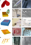
Fig. 3. Evolution of form factors for neural interfacing.
(A to C) Rigid, Si-based probes are commercially available and considered state of the art. (B) Si-based Michigan probe, modified with a patterned waveguide. Reproduced with permission from Son et al. (224). (C) Utah array (Blackrock Microsystems LLC). (D to F) Thick fiber and polymer-based probes. (E) Thermal drawing of macroscale preforms allows for multifunctional fibers that can bend and flex. A single fiber can contain electrical recording sites (CPE or Sn), guide light, or pass fluid. Reproduced with permission from Canales et al. (55). (F) Polymer probes (based on PI and SU-8) can also be assembled to support optical and fluidic stimulation and electrical interfacing. Reproduced with permission from Rubehn et al. (56). (G to I) Elastomeric probes are generally thick but are compliant and stretchable. (H) PDMS probe with off-the-shelf components and serpentine metallic structures. Reproduced with permission from Park et al. (59). (I) EDura: PDMS-based probe with electrodes and a microfluidic. Reproduced with permission from Minev et al. (58). (J to L) Ultrathin arrays and probes. (K) Neurogrid array: PEDOT:PSS-coated Au electrode sites on 4-μm PaC. Reproduced with permission from Khodagholy et al. (67). (L) SU-8 and Au array on silk fibroin that can be dissolved away to leave a mesh. Reproduced with permission from Kim et al. (68). (M to O) Freestanding mesh probes. (N) Stressed struts allow for global scrolling to form a probe-like geometry or (O) meshes that can be injected through a syringe. Reproduced with permission from Liu et al. (74) and Xie et al. (75). The colors used in the schematics on the left correspond roughly to the Young’s modulus scale in Fig. 2A.
Fig. 4
Fig. 4. Transient, bioresorbable electronics.
(A) Transient bioelectronic ECoG array micrographs of active ECoG array with Si transistors under accelerated (high pH) testing conditions. (B) Recording from three channels and a control (nonbioresorbable channel) over 33 days in vivo. The transient array is fully functional for >30 days in vivo. Reproduced with permission from Yu et al. (72).
Fig. 5
Fig. 5. Optical neural recording strategies.
Diverse approaches use the neuron activity–dependent modulation of light absorption or emission to monitor single-cell or population activity. (A) Voltage-sensitive dyes (for VSDI) accumulate within the membranes of neurons and change their conformation depending on the membrane potential, leading to changes in light emission. (B) Intrinsic imaging monitors absorption and emission of wavelengths that correlate to those of metabolic biomolecules whose numbers or composition depends on neural activity. (C) GEVIs are engineered proteins that consist of a voltage-dependent domain that is embedded in the neuron membrane and a fluorescent protein. Membrane potential changes the conformation of the protein, thereby changing fluorescence emission. (D) GECIs are engineered proteins that have a calcium-binding domain (blue) connecting a fluorescent indicator (loop). Protein conformation changes with calcium binding, leading to a change in fluorescent emission. Both (C) and (D) are genetically encoded.
Fig. 6
Fig. 6. Schematics of optical stimulation methods.
(A) Two-photon uncaging uses biologically inert neurotransmitters that become available for receptor binding after their caged moiety absorbs photons of the correct wavelength, thereby giving researchers the ability to temporally and anatomically control receptor activation. UV, ultraviolet. (B) Optogenetic stimulation with genetically encoded light-sensitive ion channel (for example, channelrhodopsin). (C) Upconverting neural stimulation by upconverting nanoparticles acting on light-sensitive ion channels. (D) Photoelectric stimulation with semiconducting devices or particles acting on voltage-sensitive ion channels. (E) Photothermal stimulation with metallic (for example, gold) nanoparticles acting on cell membrane or heat-sensitive channels.
Fig. 7
Fig. 7. Schematic of suggested cell-specific magnetic stimulation methods.
(A and C) Magnetomechanical stimulation with superparamagnetic nanoparticles or magnetic proteins acting on a mechanosensitive channel (for example, TREK1). (B and D) Magnetothermal stimulation with superparamagnetic nanoparticles or magnetic proteins acting on a heat-sensitive channel (for example, TRPV1). The magnetic nature of the protein-based stimulation approaches (C and D), that is, coexpression of ferritin with the mechanosensitive/heat-sensitive ion channels, is currently debated and requires further investigation and confirmation of the proposed underlying mechanisms.
Fig. 8
Fig. 8. Schematic of suggested ultrasonic stimulation methods.
(A) Ultrasonic stimulation with microbubbles acting on mechanosensitive channels (that is, TRP-4). (B) Ultrasonic stimulation with piezoelectric nanoparticles acting on voltage-sensitive channels.

References

    1. Deep-Brain Stimulation for Parkinson’s Disease Study Group, Deep-brain stimulation of the subthalamic nucleus or the pars interna of the globus pallidus in Parkinson’s disease. N. Engl. J. Med. 345, 956–963 (2001). - PubMed
    1. Ondo W., Jankovic J., Schwartz K., Almaguer M., Simpson R. K., Unilateral thalamic deep brain stimulation for refractory essential tremor and Parkinson’s disease tremor. Neurology 51, 1063–1069 (1998). - PubMed
    1. Greenberg B. D., Malone D. A., Friehs G. M., Rezai A. R., Kubu C. S., Malloy P. F., Salloway S. P., Okun M. S., Goodman W. K., Rasmussen S. A., Three-year outcomes in deep brain stimulation for highly resistant obsessive–compulsive disorder. Neuropsychopharmacology 31, 2384–2393 (2006). - PubMed
    1. Mayberg H. S., Lozano A. M., Voon V., McNeely H. E., Seminowicz D., Hamani C., Schwalb J. M., Kennedy S. H., Deep brain stimulation for treatment-resistant depression. Neuron 45, 651–660 (2005). - PubMed
    1. Bouton C. E., Shaikhouni A., Annetta N. V., Bockbrader M. A., Friedenberg D. A., Nielson D. M., Sharma G., Sederberg P. B., Glenn B. C., Mysiw W. J., Morgan A. G., Deogaonkar M., Rezai A. R., Restoring cortical control of functional movement in a human with quadriplegia. Nature 533, 247–250 (2016). - PubMed

Publication types