. 2017 Jun 7;3(6):e1700147.
doi: 10.1126/sciadv.1700147. eCollection 2017 Jun.

Ribosome rearrangements at the onset of translational bypassing

Affiliations

Ribosome rearrangements at the onset of translational bypassing

Xabier Agirrezabala et al. Sci Adv. .

Abstract

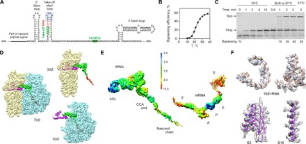
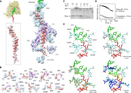
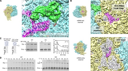
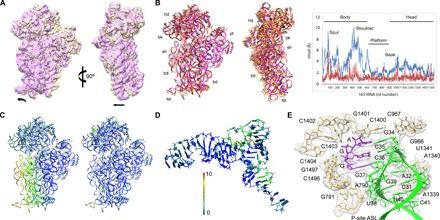
Bypassing is a recoding event that leads to the translation of two distal open reading frames into a single polypeptide chain. We present the structure of a translating ribosome stalled at the bypassing take-off site of gene 60 of bacteriophage T4. The nascent peptide in the exit tunnel anchors the P-site peptidyl-tRNAGly to the ribosome and locks an inactive conformation of the peptidyl transferase center (PTC). The mRNA forms a short dynamic hairpin in the decoding site. The ribosomal subunits adopt a rolling conformation in which the rotation of the small subunit around its long axis causes the opening of the A-site region. Together, PTC conformation and mRNA structure safeguard against premature termination and read-through of the stop codon and reconfigure the ribosome to a state poised for take-off and sliding along the noncoding mRNA gap.

Keywords: bypassing; recoding; ribosome; structure; translation.


Figures

Fig. 1
Fig. 1. Ribosome stalling at a take-off codon upon translational bypassing on bacteriophage T4 gene 60.
(A) Key regulatory elements in gene 60 mRNA, including the nascent peptide and the mRNA SL elements. The apical part of the take-off SL is highlighted in gray. (B) Temperature dependence of bypassing. (C) Reversibility of the temperature-induced ribosome stalling at the take-off codon. When translation is carried out at 10°C, only the stop peptide corresponding to codons 1 to 46 is synthesized. Switching the temperature to 37°C activates bypassing (Byp product). The last lane shows the maximum bypassing efficiency when translation is performed at 37°C. (D) Cryo-EM densities showing 30S (yellow) and 50S (blue) subunits, the P-site tRNAGly (green) carrying the nascent peptide (red), and the mRNA (magenta) (see also fig. S1). (E) Local resolution. The scale bar shows a color scale with resolution in angstroms (see also figs. S2 and S3). ASL, anticodon stem-loop. (F) Examples of the experimental cryo-EM density (transparent gray) and atomic model (ribbons), including 16S rRNA (nucleotides 766 to 779 and 586 to 639) and proteins S3 and S15 (residues 110 to 170 and 48 to 72, respectively).
Fig. 2
Fig. 2. The peptide exit tunnel and the PTC.
(A) Nascent chain interactions in the exit tunnel. The cryo-EM density is presented in transparent gray (peptide) and blue/purple (the 50S subunit 23S rRNA and proteins, respectively). The peptide density is shown at a lower contour level. (B) Closeup view of the interactions. The cryo-EM density is shown in transparent gray. (C) RF1 is inactive on the take-off complexes at low temperatures. Ribosome complexes formed in the presence of RF1 were isolated by gel filtration, which separates the complexes from free RF1 and released nascent chains. The amounts of RF1 and nascent chains copurifying in the isolated complex were analyzed by SDS–polyacrylamide gel electrophoresis (PAGE) and imaging of the fluorescence reporters on RF1 and the nascent chain. The last lane shows the control with RF1 alone. (D) Kinetics of the Pmn reaction of two peptidyl-tRNAs, one stalled before bypassing at the take-off codon and carrying the stop peptide (Take-off + Pmn; closed circles) and another carrying a shorter peptide, which appears as a transient intermediate of translation at 10°C (Translation + Pmn; open circles) (see also fig. S5). a.u., arbitrary units. (E) Conformation of the PTC in the take-off complex (upper left), compared to the induced (upper right) and uninduced (lower left) conformations, and in a complex with RF1 (lower left; the GGQ motif is shown in yellow) (53).
Fig. 3
Fig. 3. The mRNA path.
(A) mRNA hairpin in the A site. Closeup view of the mRNA (magenta), P-site peptidyl-tRNAGly (green), and the modeled sequence. Potential G-C pairs are marked by asterisks. The mRNA density is shown at a lower contour level. (B) Visualization of the mRNA densities of the 5′ and 3′ regions. Top: The 5′ region of the mRNA. The 3′ end of 16S rRNA (nucleotides 1528 to 1540) and protein L30 are highlighted. Bottom: The 3′ end of the mRNA as it emerges from the channel. Proteins S3, S4, and S5 are also highlighted. (C) Importance of the A-site hairpin for the discrimination against RF1. Left: mRNA constructs used, with the native sequence of gene 60 [wild type (WT)] or lacking one G-C base pair from the apical part of the take-off hairpin (−GC). Middle: Translation of WT and –GC mRNAs in the presence of different concentrations of RF1 at 37°C. Right: Dependence of the bypassing efficiency on the RF1 concentration. R, RF1/70S ratio under half-inhibition conditions (see also fig. S4). (D) A-site hairpin as a guard against UAG read-through (rt). Translation of the WT mRNA (left) and −GC mRNA (right) at different temperatures. Note the accumulation of the read-through product at low temperatures during translation of −GC mRNA.
Fig. 4
Fig. 4. A rolling motion of the 30S subunit.
(A) Comparison of the 30S subunit in the take-off complex (yellow) and in the classical pretranslocation state (purple; see Materials and Methods) shown as intersubunit (left) and side (right) views. The 50S subunit was used for the alignment. Arrows indicate directions of movement. (B) Global rearrangements of the 30S subunit. Left: Superposition of the take-off complex with the unrolled 70S–EF-Tu complex [Protein Data Bank (PDB) 5AFI (46)] shown as intersubunit (left) and side (right) views. Right: The graph shows the movements of 16S rRNA (backbone distances) in the take-off complex compared to the unrolled ribosome structure (blue) or the SecM-arrested complex (red) [PDB 3JBU (28)]. 23S rRNA was used as reference for alignment. rmsd, root mean square deviation. (C) The heat map displayed on the take-off complex quantifies the displacement, with respect to the ribosome with EF-Tu bound (left) and the SecM-arrested complex (right). (D) Deviations in the structure of tRNAGly compared to the 70S–EF-Tu complex. 23S rRNA was used as reference for alignment. (E) Interactions of the tRNAGly anticodon SL. The tRNA and mRNA binding the P site are stabilized by the stacking of the ribose and base of nucleotide 34 in the anticodon with G966 and C1400 in 16S rRNA, respectively, and the contacts involving the universally conserved nucleotide A790 with the tRNA nucleotide 38, nucleotides G1338 and A1339 with nucleotide 41, and the G30-C40 base pair in the tRNA, consistent with earlier structural work (32, 33). The cryo-EM density is shown in transparent gray.

References

    1. Baranov P. V., Atkins J. F., Yordanova M. M., Augmented genetic decoding: Global, local and temporal alterations of decoding processes and codon meaning. Nat. Rev. Genet. 16, 517–529 (2015). - PubMed
    1. Lang B. F., Jakubkova M., Hegedusova E., Daoud R., Forget L., Brejova B., Vinar T., Kosa P., Fricova D., Nebohacova M., Griac P., Tomaska L., Burger G., Nosek J., Massive programmed translational jumping in mitochondria. Proc. Natl. Acad. Sci. U.S.A. 111, 5926–5931 (2014). - PMC - PubMed
    1. Huang W. M., Ao S. Z., Casjens S., Orlandi R., Zeikus R., Weiss R., Winge D., Fang M., A persistent untranslated sequence within bacteriophage T4 DNA topoisomerase gene 60. Science 239, 1005–1012 (1988). - PubMed
    1. Weiss R. B., Huang W. M., Dunn D. M., A nascent peptide is required for ribosomal bypass of the coding gap in bacteriophage T4 gene 60. Cell 62, 117–126 (1990). - PMC - PubMed
    1. Herr A. J., Gesteland R. F., Atkins J. F., One protein from two open reading frames: Mechanism of a 50 nt translational bypass. EMBO J. 19, 2671–2680 (2000). - PMC - PubMed

Publication types

LinkOut - more resources