Multivariate classification of pain-evoked brain activity in temporomandibular disorder
- PMID: 28630949
- PMCID: PMC5473632
- DOI: 10.1097/PR9.0000000000000572
Multivariate classification of pain-evoked brain activity in temporomandibular disorder
Abstract
Introduction: Central nervous system factors are now understood to be important in the etiology of temporomandibular disorders (TMD), but knowledge concerning objective markers of central pathophysiology in TMD is lacking. Multivariate analysis techniques like support vector machines (SVMs) could generate important discoveries regarding the expression of pain centralization in TMD. Support vector machines can recognize patterns in “training” data and subsequently classify or predict new “test” data.
Objectives: We set out to detect the presence and location of experimental pressure pain and determine clinical status by applying SVMs to pain-evoked brain activity.
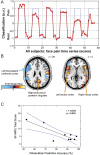
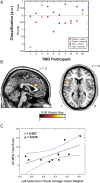
Methods: Functional magnetic resonance imaging was used to record brain activity evoked by subjectively equated noxious temporalis pressures in patients with TMD and controls. First, we trained an SVM to recognize when the evoked pain stimulus was on or off based on each individual's pain-evoked blood–oxygen–level–dependent (BOLD) signals. Next, an SVM was trained to distinguish between the BOLD response to temporalis-evoked pain vs thumb-evoked pain. Finally, an SVM attempted to determine clinical status based on temporalis-evoked BOLD.
Results: The on-versus-off accuracy in controls and patients was 83.3% and 85.1%, respectively, both significantly better than chance (ie, 50%). Accurate determination of experimental pain location was possible in patients with TMD (75%), but not in healthy subjects (55%). The determination of clinical status with temporalis-evoked BOLD (60%) failed to reach statistical significance.
Conclusion: The SVM accurately detected the presence of noxious temporalis pressure in patients with TMD despite the stimulus being colocalized with their ongoing clinical pain. The SVM's ability to determine the location of noxious pressure only in patients with TMD reveals somatotopic-dependent differences in central pain processing that could reflect regional variations in pain valuation.
Keywords: Artificial intelligence; Brain function; Magnetic resonance imaging (MRI); Neuroscience/Neurobiology; Orofacial pain/TMD; Support vector machines (SVM).
Conflict of interest statement
Sponsorships or competing interests that may be relevant to content are disclosed at the end of this article.
Figures


References
-
- Barros Vde M, Seraidarian PI, Cortes MI, de Paula LV. The impact of orofacial pain on the quality of life of patients with temporomandibular disorder. J Orofac Pain 2009;23:28–37. - PubMed
Grants and funding
LinkOut - more resources
Full Text Sources
Other Literature Sources
