Review
. 2018 Feb;93(1):350-363.
doi: 10.1111/brv.12345. Epub 2017 Jun 19.

Osteoblast migration in vertebrate bone

Affiliations
Review

Osteoblast migration in vertebrate bone

Antonia Thiel et al. Biol Rev Camb Philos Soc. 2018 Feb.

Abstract

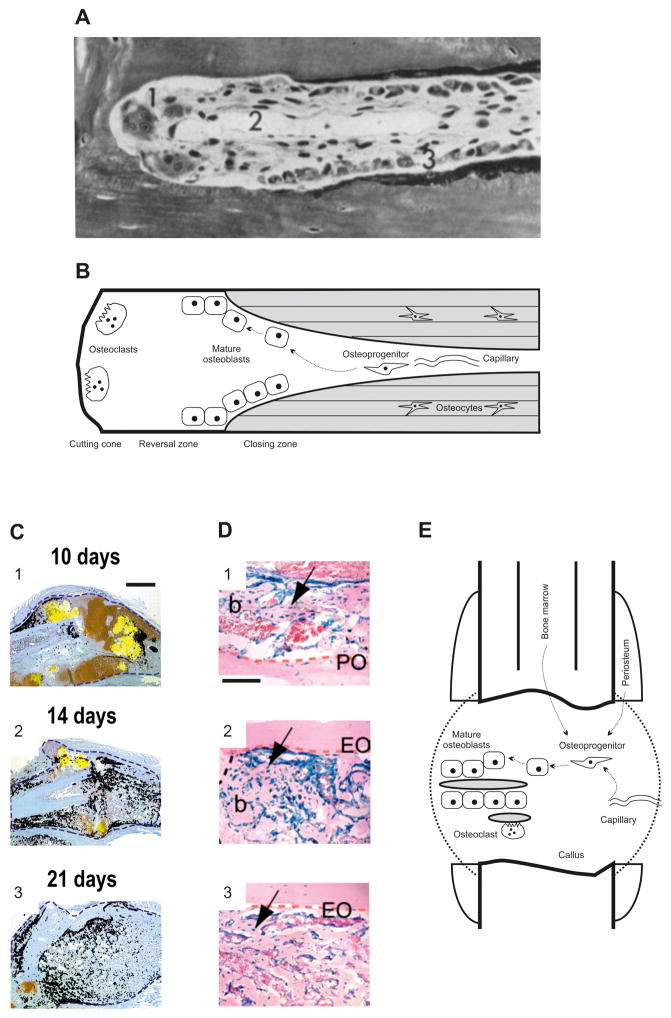
Bone formation, for example during bone remodelling or fracture repair, requires mature osteoblasts to deposit bone with remarkable spatial precision. As osteoblast precursors derive either from circulation or resident stem cell pools, they and their progeny are required to migrate within the three-dimensional bone space and to navigate to their destination, i.e. to the site of bone formation. An understanding of this process is emerging based on in vitro and in vivo studies of several vertebrate species. Receptors on the osteoblast surface mediate cell adhesion and polarization, which induces osteoblast migration. Osteoblast migration is then facilitated along gradients of chemoattractants. The latter are secreted or released proteolytically by several cell types interacting with osteoblasts, including osteoclasts and vascular endothelial cells. The positions of these cellular sources of chemoattractants in relation to the position of the osteoblasts provide the migrating osteoblasts with tracks to their destination, and osteoblasts possess the means to follow a track marked by multiple chemoattractant gradients. In addition to chemotactic cues, osteoblasts sense other classes of signals and utilize them as landmarks for navigation. The composition of the osseous surface guides adhesion and hence migration efficiency and can also provide steering through haptotaxis. Further, it is likely that signals received from surface interactions modulate chemotaxis. Besides the nature of the surface, mechanical signals such as fluid flow may also serve as navigation signals for osteoblasts. Alterations in osteoblast migration and navigation might play a role in metabolic bone diseases such as osteoporosis.

Keywords: bone; cell migration; chemotaxis; fluid flow; mineralized surfaces; osteoblasts.


Figures

Fig. 1
Fig. 1
Bone formation and the requirement for osteoblast migration in bone remodelling and endochondral fracture healing. (A) Goldner stain of a cortical basic morphometric unit (BMU) in dog radius. 1, osteoclasts; 2, central capillary; 3, osteoblasts. Reprinted with permission from (Schenk & Willenegger, 1964). Magnification 320:1. (B) Model of the structure and cellular organization of a cortical BMU. Schematic derived from histological assessments such as that in A. Newly formed bone is shaded grey. The dashed arrows denote potential migration paths of osteoblasts. (C) Closed, non-stabilized tibial fracture in mouse. Transcripts for the osteoblast marker osteocalcin (black) and the hypertrophic chondrocyte marker collagen type X (yellow) were superimposed onto adjacent tissue sections stained with safranin-O/fast green. 1, on day 10, osteoblasts deposit bone mainly on the periosteal surface. 2, day 14 is characterized by a large number of individual areas of bone formation throughout the entire callus. 3, after 21 days, osteoblast activity remains prominent in the callus. Blue dotted lines represent the boundary of the callus. Scale bar for images 1–3, 1 mm. Reprinted with permission from Colnot (2003). (D) Bone grafts derived from lacZ reporter gene mice were used to track cells during healing of non-stabilized tibial fractures. The following three tissue sources were tracked: 1, periosteum; 2, endosteum; 3, endosteum and bone marrow. Images show X-gal stainings on day 14. Osteoblasts were found to derive from all three tissue sources. Abbreviations: b, bone; EO, endosteal surface; PO, periosteal surface. The arrow denotes graft-derived osteoblasts/osteocytes. Dotted orange and black lines delimit the bone graft and bone/cartilage junction, respectively. Scale bar for images 1–3: 100 μm. Reprinted with permission from Colnot et al. (2009). (E) Model of the structure and cellular organization of a typical fracture callus. Representative areas of newly formed bone are shaded grey. The dashed arrows denote potential migration paths of osteoblasts.
Fig. 2
Fig. 2
Osteoblast migration. (A) The osteoblast layer on the endocranial aspect of rat parietal bones was mechanically disrupted and the subsequent migration of osteoblasts onto the cleared bone matrix observed using scanning electron microscopy (SEM) at several time points. 1, SEM at 420 μm field width acquired 8 h post cell clearance. Osteoblasts had partially repopulated the cleared surface. The first-rank cells, i.e. the migration front (marked by arrow) had moved away from their neighbours so that their cell bodies were no longer close to other osteoblasts, although other osteoblasts migrated in a densely packed formation. 2, SEM at 160 μm field width acquired 24 h post cell clearance. Some of the spaced cells of the most forward ranks had migrated with apparent disregard for the pattern of the collagen they had traversed. Reprinted with permission from Jones & Boyde (1977). (B) Dynamic, non-invasive molecular imaging of osteoblast migration in vivo. Still images from confocal time-lapse microscopy of migrating photoconverted Kaede-labelled zebrafish (Danio rerio) osteoblasts (red) in close proximity to the fracture. Protrusions of one cell are highlighted in yellow, its leading edge with an arrowhead. Abbreviations: BF, bright field; dpi, day post injury; entpd5, ectonucleoside triphosphate diphosphohydrolase 5. Scale bar, 20 μm. Reprinted with permission from Geurtzen et al. (2014).
Fig. 3
Fig. 3
Topography recognition of osteoblasts. Scanning electron microscope (SEM) images of osteoblasts on experimental substrates with a fixed groove depth of 330 nm. Cells were immunolabelled using an antibody against vinculin, a marker of adhesion complexes. (A) Osteoblasts on 25 μm grooves formed adhesions predominantly on the raised ridge areas (indicated in insert). (B) Diffuse 100 μm grooves did not affect adhesion formation. Scale bars are shown. Reprinted with permission from Biggs et al. (2008).
Fig. 4
Fig. 4
Working model osteoblast navigation. Osteoprogenitor cells originate from vascular structures or stem cell pools. The migration direction of movement of osteoprogenitors and their progeny is governed by gradients of chemoattractants (shaded grey) which are released either proteolytically from matrix through osteoclastic resorption or are secreted by neighbouring cell types. Osteoblasts can also sense mechanical signals, for example from fluid flow, that can guide the tracks of osteoblasts. Generally, migration of osteoblasts occurs on surfaces such as resorbed bone or newly formed osteoid and these surfaces control, for example, cell adhesion or receptors for chemotaxis; they can also provide substrate-bound chemoattractants for haptotaxis. Abbreviations: IL-1β, interleukin β1; TGF-β1, transforming growth factor-β1.

References

    1. Abdelgawad ME, Soe K, Andersen TL, Merrild DM, Christiansen P, Kjaersgaard-Andersen P, Delaisse JM. Does collagen trigger the recruitment of osteoblasts into vacated bone resorption lacunae during bone remodeling? Bone. 2014;67:181–188. - PubMed
    1. Baldini G, Ponti C, Bortul R, Narducci P, Grill V, Martelli AM. Sparc localizes to the blebs of hobit cells and human primary osteoblasts. Journal of Cellular Biochemistry. 2008;104:2310–2323. - PubMed
    1. Balogh ZJ, Reumann MK, Gruen RL, Mayer-Kuckuk P, Schuetz MA, Harris IA, Gabbe BJ, Bhandari M. Advances and future directions for management of trauma patients with musculoskeletal injuries. Lancet. 2012;380:1109–1119. - PubMed
    1. Basso FG, Silveira Turrioni AP, Hebling J, de Souza Costa CA. Zoledronic acid inhibits human osteoblast activities. Gerontology. 2013;59:534–541. - PubMed
    1. Baum B, Settleman J, Quinlan MP. Transitions between epithelial and mesenchymal states in development and disease. Seminars in Cell & Developmental Biology. 2008;19:294–308. - PubMed

Publication types