. 2017 Jun;98(6):1282-1293.
doi: 10.1099/jgv.0.000815. Epub 2017 Jun 20.

Loss of Sendai virus C protein leads to accumulation of RIG-I immunostimulatory defective interfering RNA

Affiliations

Loss of Sendai virus C protein leads to accumulation of RIG-I immunostimulatory defective interfering RNA

Maria Teresa Sánchez-Aparicio et al. J Gen Virol. 2017 Jun.

Abstract

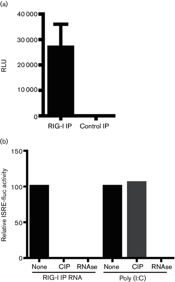
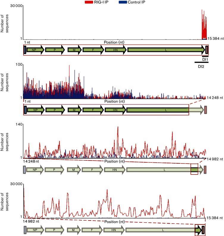
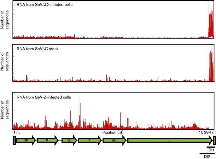
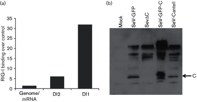
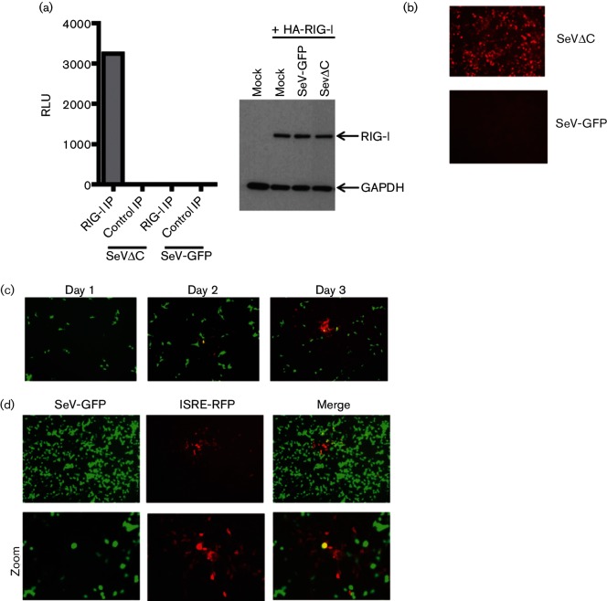
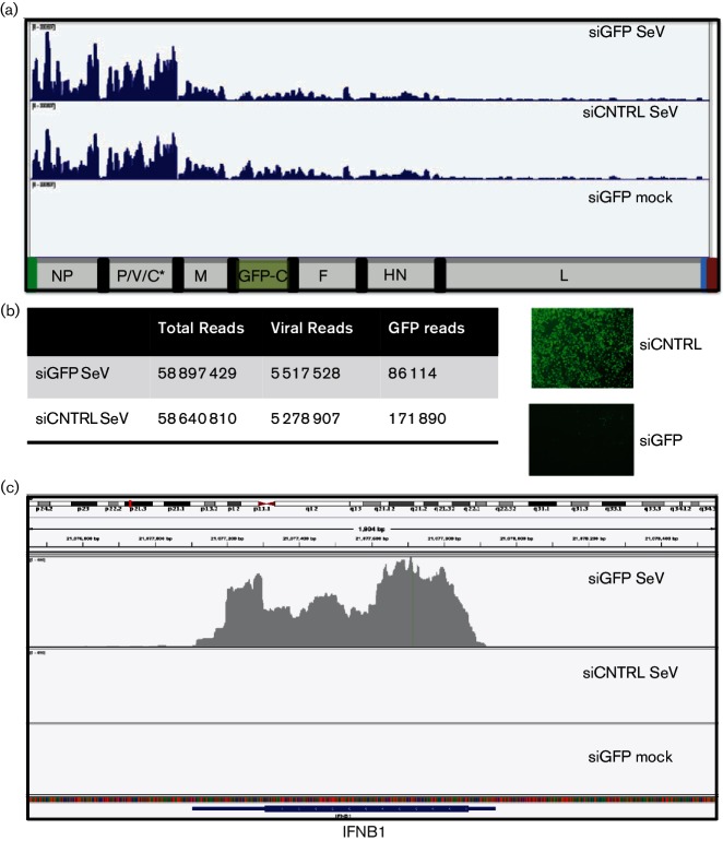
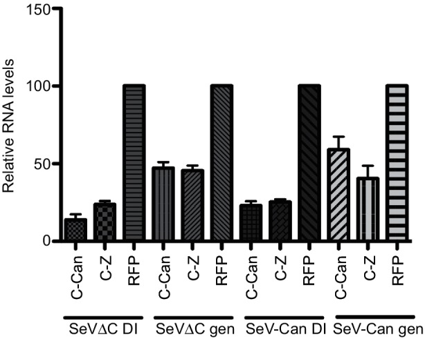
Retinoic acid inducible gene (RIG-I)-mediated innate immunity plays a pivotal role in defence against virus infections. Previously we have shown that Sendai virus (SeV) defective interfering (DI) RNA functions as an exclusive and potent RIG-I ligand in DI-RNA-rich SeV-Cantell infected cells. To further understand how RIG-I is activated during SeV infection, we used a different interferon (IFN)-inducing SeV strain, recombinant SeVΔC, which, in contrast to SeV-Cantell is believed to stimulate IFN production due to the lack of the SeV IFN antagonist protein C. Surprisingly, we found that in SevΔC-infected cells, DI RNAs also functioned as an exclusive RIG-I ligand. Infections with wild-type SeV failed to generate any RIG-I-associated immunostimulatory RNA and this correlated with the lack of DI genomes in infected cells, as well as with the absence of cellular innate immune responses. Supplementation of the C protein in the context of SeVΔC infection led to a reduction in the number of DI RNAs, further supporting the potential role of the C protein as a negative regulator of DI generation and/or accumulation. Our findings indicate that limiting DI genome production is an important function of viral IFN antagonist proteins.


Conflict of interest statement

The authors declare that there are no conflicts of interest.

Figures

Fig. 1.
Fig. 1.
Immunostimulatory activity of RIG-I-associated RNA. (a) RIG-I-associated RNA from SeVΔC-infected A549 cells or control IP RNA was transfected into 293T ISRE-Fluc cells. Data from three individual experiments with SD are shown. (b) ISRE-Fluc reporter activation by RIG-I IP RNA or poly(I:C) treated with calf alkaline phosphatase (CI) and RNAse A.
Fig. 2.
Fig. 2.
SeVΔC RNAs enriched in RIG-I IPs. Total RNA isolated with RIG-I (red) or control (blue) IP was subjected to deep sequencing. RNA-Seq reads are mapped to their corresponding position within the SeV genome. The top panel illustrates the entire SeV genome. The bottom three panels zoom in on highlighted regions of the SeV genome. The number of RNA-Seq reads is given on the Y-axis. RIG-I-specific enrichment, corresponding to DI RNAs, can be seen in the bottom two panels. The bottom panel encompasses reads mapping to both DI1 and DI2. The second from the bottom panel corresponds to reads mapping to DI2.
Fig. 3.
Fig. 3.
RNA-Seq profiles of SeVΔC-infected cells and virus stock. Reads from RNA-Seq were mapped to the SeV genome. DI RNA can be visualized as a strong peak on the 5′ end of the genome in both RNA from SeVC-infected cells and RNA from purified virus stock (top two panels). No DI RNA is apparent in SeV-Z-infected cells (bottom panel).
Fig. 4.
Fig. 4.
(a) Quantitative enrichment of RIG-I binding by SeV RNA species. Ratios of RIG-I-associated reads over control IP reads were calculated. (b) 293 T cells were infected with the various Sendai viruses and total cell lysates were analysed by Western blot with SeV C protein antiserum 24 h post-infection. Bands corresponding to C protein, as well as P and V proteins, can be seen.
Fig. 5.
Fig. 5.
DI-free SeV is a weak activator of ISRE reporter. (a) Immunostimulatory activity of HA-RIG-I associated RNA from 293 T cells infected with either SeVΔC or SeV-GFP. In the right panel, a Western blot shows the expression levels of HA-RIG-I in both infections. (b) 293T ISRE-RFP reporter cells were infected with either SeVΔC or SeV-GFP and infections were allowed to proceed for 24 h. Red fluorescent protein (RFP) expression correlates to the immunostimulatory potential of each virus. (c) Infection of ISRE-RFP cells with SeV-GFP results in minimal RFP expression isolated to a few cells over the first 72 h of infection. (d) Individual RFP plaques surrounding SeV-GFP-infected cells at 96h post-infection. The bottom panel is a magnified view of the images in the top panel. The single yellow cell in the middle of the merged image represents a double-positive cell.
Fig. 6.
Fig. 6.
Knockdown of SeV C protein in infected cells. HeLa cells were transfected with siGFP or siCNTRL and infected with SeV-GFP-C (which expresses a GFP-C fusion protein). (a) Sendai RNA-Seq reads from siRNA-transfected and SeV- or mock-infected cells. The top two panels highlight very similar viral profiles in the presence or absence of C protein. (b) Comparison of loss of GFP RNA versus GFP protein, illustrating that the majority of the GFP protein is removed in siGFP cells. (c) RNA-Seq reads from siRNA-treated and infected cells were mapped to the human IFNB gene. Very strong IFNB expression can be seen in infected cells with GFP-C knockdown.
Fig. 7.
Fig. 7.
Exogenous C protein limits production of cbDI as well as genomic RNA. Relative expression of SeV cpDI or genomic RNA in the presence or absence of SeV C protein. SeV-Can or SeV-Z C protein-expressing plasmids were transfected into HeLa cells subsequently infected with either SeVΔC or SeV-Cantell. Viral RNA was measured at 24 h post-infection by q-RT-qPCR. Viral RNA levels in RFP-transfected control cells were set to 100 %. Normalized data from three individual experiments are shown with sd.

Similar articles

Cited by

References

    1. Janeway CA, Medzhitov R. Innate immune recognition. Annu Rev Immunol. 2002;20:197–216. doi: 10.1146/annurev.immunol.20.083001.084359. - DOI - PubMed
    1. Baum A, García-Sastre A. Induction of type I interferon by RNA viruses: cellular receptors and their substrates. Amino Acids. 2010;38:1283–1299. doi: 10.1007/s00726-009-0374-0. - DOI - PMC - PubMed
    1. Hornung V, Ellegast J, Kim S, Brzózka K, Jung A, et al. 5′-Triphosphate RNA is the ligand for RIG-I. Science. 2006;314:994–997. doi: 10.1126/science.1132505. - DOI - PubMed
    1. Pichlmair A, Schulz O, Tan CP, Näslund TI, Liljeström P, et al. RIG-I-mediated antiviral responses to single-stranded RNA bearing 5′-phosphates. Science. 2006;314:997–1001. doi: 10.1126/science.1132998. - DOI - PubMed
    1. Schlee M, Roth A, Hornung V, Hagmann CA, Wimmenauer V, et al. Recognition of 5′ triphosphate by RIG-I helicase requires short blunt double-stranded RNA as contained in panhandle of negative-strand virus. Immunity. 2009;31:25–34. doi: 10.1016/j.immuni.2009.05.008. - DOI - PMC - PubMed

LinkOut - more resources