. 2017 Jun 20;8(6):166.
doi: 10.3390/genes8060166.

A Comparative Study of the Structural Dynamics of Four Terminal Uridylyl Transferases

Affiliations

A Comparative Study of the Structural Dynamics of Four Terminal Uridylyl Transferases

Kevin J Cheng et al. Genes (Basel). .

Abstract

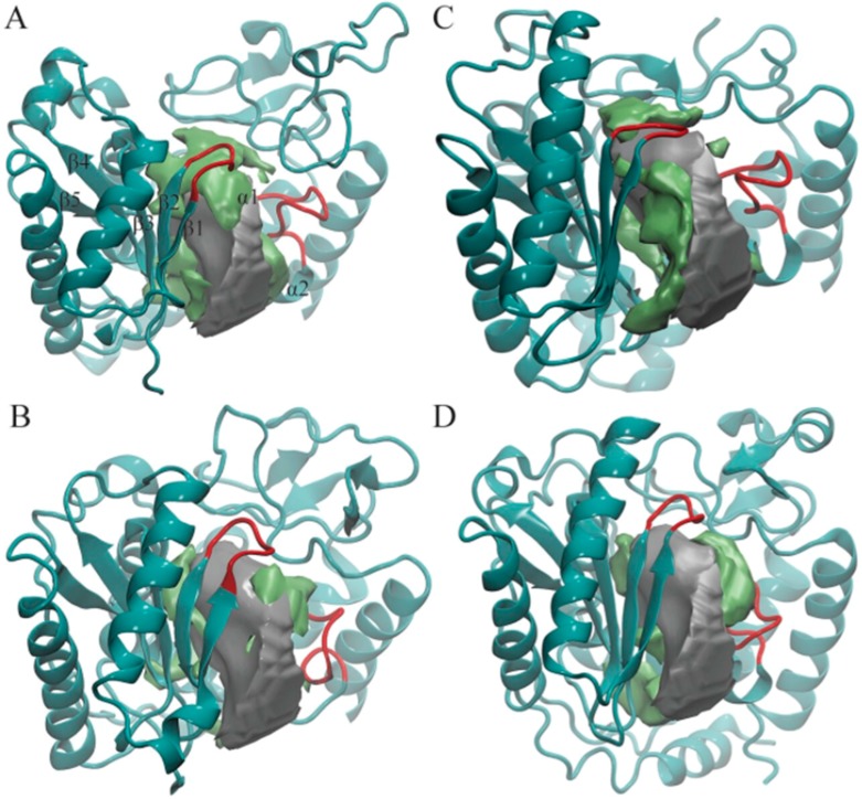
African trypanosomiasis occurs in 36 countries in sub-Saharan Africa with 10,000 reported cases annually. No definitive remedy is currently available and if left untreated, the disease becomes fatal. Structural and biochemical studies of trypanosomal terminal uridylyl transferases (TUTases) demonstrated their functional role in extensive uridylate insertion/deletion of RNA. Trypanosoma brucei RNA Editing TUTase 1 (TbRET1) is involved in guide RNA 3' end uridylation and maturation, while TbRET2 is responsible for U-insertion at RNA editing sites. Two additional TUTases called TbMEAT1 and TbTUT4 have also been reported to share similar function. TbRET1 and TbRET2 are essential enzymes for the parasite viability making them potential drug targets. For this study, we clustered molecular dynamics (MD) trajectories of four TUTases based on active site shape measured by Pocket Volume Measurer (POVME) program. Among the four TUTases, TbRET1 exhibited the largest average pocket volume, while TbMEAT1's and TbTUT4's active sites displayed the most flexibility. A side pocket was also identified within the active site in all TUTases with TbRET1 having the most pronounced. Our results indicate that TbRET1's larger side pocket can be exploited to achieve selective inhibitor design as FTMap identifies it as a druggable pocket.

Keywords: MEAT1; POVME; RET1; RET2; TUT4; TUTases; Trypanosoma brucei; electrostatics; pocket shape; pocket volume; terminal uridylyl transferases.


Conflict of interest statement

Rommie E. Amaro is a co-founder of, on the scientific advisory board of, and has equity interest in Actavalon, Inc.

Figures

Figure 1
Figure 1
Structures of four T. brucei terminal uridylyl transferases (TUTases) shown in ribbons. (A) TbRET1; (B) TbRET2; (C) TbMEAT1; (D) TbTUT4. Enzymes are colored according to the domains: C-terminal domain (CTD) in green, N-terminal domain (NTD) in purple, middle domain (MiD) (RRM domain in TbRET1) in light blue, bridge domain (BD) in dark blue, and the zinc finger domain and connecting helix of TbRET1 in silver. uridine triphosphate (UTP) is shown in orange sticks, and Zn is shown as a red sphere.
Figure 2
Figure 2
Pocket shapes of the most-populated four cluster centroids: (A) TbRET1; (B) TbRET2; (C) TbMEAT1; (D) TbTUT4. Only NTD and CTD are shown in cyan ribbons in each enzyme for clarity. The average pocket shape of all enzymes is shown as a silver surface, and the additional region opening only in that cluster are shown as a green surface. The two loops we found to modulate the pocket shape are highlighted with red. Beta-sheets and alpha-helices close to the active site are labeled on TbRET1 only (α1 labeled, but not visible).
Figure 3
Figure 3
Ensemble-averaged electrostatics of (A) TbRET1; (B) TbRET2; (C) TbMEAT1; (D) TbTUT4. The exact same orientations shown in Figure 1 are used to demonstrate the electrostatic potential surface on the left, and the rear views are shown on the right. The blue patches represent areas of positive electrostatic potential, and the red patches represent areas of negative potential. Active site is designated with a dashed oval.
Figure 4
Figure 4
A side pocket was identified from visualizing our POVME results for the centroid of Cluster 1 (TbRET1, Panel A). The same region is compared with the centroid of Cluster 2 (TbRET2 Panel B) to demonstrate the pronounced pocket volume increase. Additional regions opening only in that cluster are shown as a green surface relative to the average pocket volume. The side pocket is located between β4 and β5.

References

    1. Berriman M., Ghedin E., Hertz-Fowler C., Blandin G., Renauld H., Bartholomeu D.C., Lennard N.J., Caler E., Hamlin N.E., Haas B., et al. The genome of the african trypanosome trypanosoma brucei. Science. 2005;309:416–422. doi: 10.1126/science.1112642. - DOI - PubMed
    1. Demir O., Amaro R.E. Elements of nucleotide specificity in the trypanosoma brucei mitochondrial RNA editing enzyme RET2. J. Chem. Inf. Model. 2012;52:1308–1318. doi: 10.1021/ci3001327. - DOI - PMC - PubMed
    1. Buchanan-Kilbey G., Djumpah J., Papadopoulou M.V., Bloomer W., Hu L., Wilkinson S.R., Ashworth R. Evaluating the developmental toxicity of trypanocidal nitroaromatic compounds on zebrafish. Acta Trop. 2013;128:701–705. doi: 10.1016/j.actatropica.2013.07.022. - DOI - PubMed
    1. Ganesan A., Coote M.L., Barakat K. Molecular dynamics-driven drug discovery: Leaping forward with confidence. Drug Discov. Today. 2017;22:249–269. doi: 10.1016/j.drudis.2016.11.001. - DOI - PubMed
    1. Deng J., Ernst N.L., Turley S., Stuart K.D., Hol W.G. Structural basis for UTP specificity of RNA editing tutases from trypanosoma brucei. EMBO J. 2005;24:4007–4017. doi: 10.1038/sj.emboj.7600861. - DOI - PMC - PubMed