

Fluorescence Analysis of Vitamin D Receptor Status of Circulating Tumor Cells (CTCS) in Breast Cancer: From Cell Models to Metastatic Patients
- PMID: 28632174
- PMCID: PMC5486139
- DOI: 10.3390/ijms18061318
Fluorescence Analysis of Vitamin D Receptor Status of Circulating Tumor Cells (CTCS) in Breast Cancer: From Cell Models to Metastatic Patients
Abstract
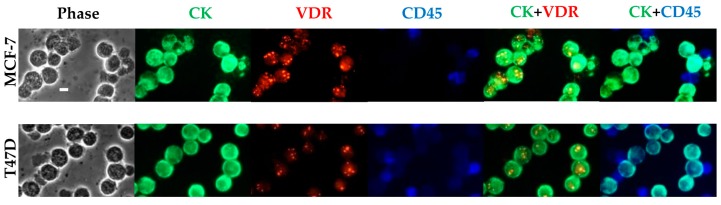
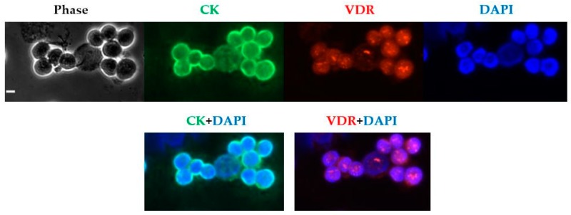
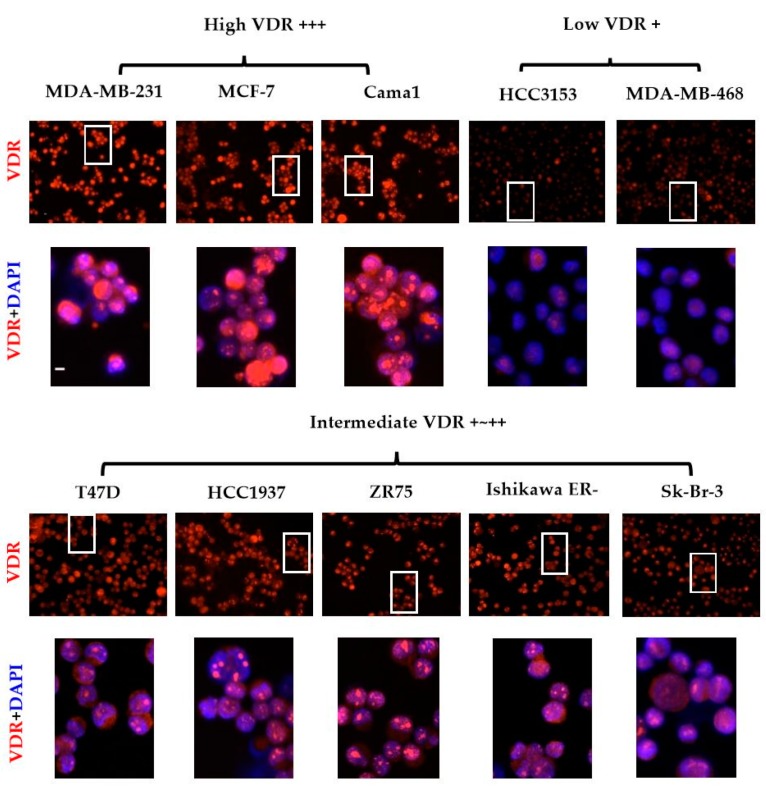
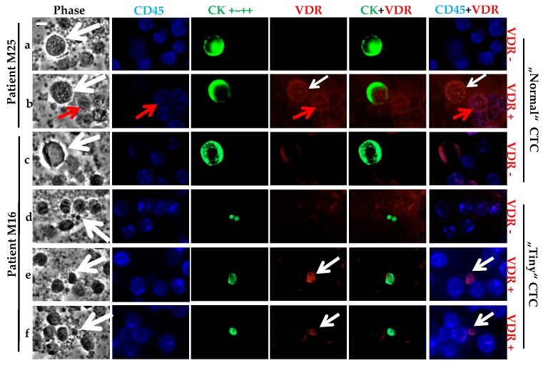
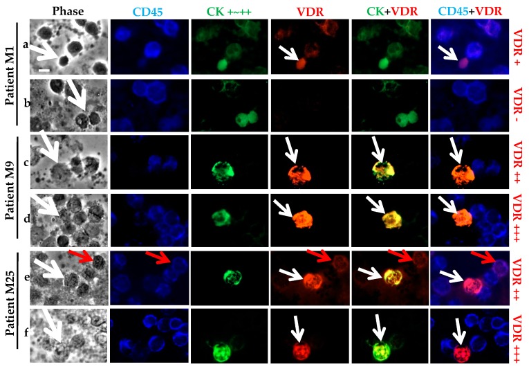
The Vitamin D receptor (VDR) expressed in normal breast tissue and breast tumors has been suggested as a new prognostic biomarker in breast cancer (BC). Besides, increasing evidence supports the view that the detection of circulating tumor cells (CTCs) predicts outcome in early and metastatic BC. Consequently, an evaluation of VDR expression in the CTCs of BC patients may allow optimization of their treatment. As an attempt to profile and subtype the CTCs of metastatic patients, we established an innovative fluorescence technique using nine BC cell lines to visualize, define, and compare their individual VDR status. Afterwards, we tested the CTC presence and VDR expression in blood samples (cytospins) collected from 23 metastatic BC patients. The results demonstrated major differences in the VDR levels among the nine cell lines, and VDR positive CTCs were detected in 46% of CTC-positive patients, with a total of 42 CTCs individually analyzed. Due to the limited number of patients in this study, no correlation between VDR expression and BC subtype classification (according to estrogen receptor (ER), progesterone receptor (PR) and HER2) could be determined, but our data support the view that VDR evaluation is a potential new prognostic biomarker to help in the optimization of therapy management for BC patients.
Keywords: breast cancer; circulating tumor cells; vitamin D receptor.
Conflict of interest statement
The authors declare no conflict of interest.
Figures
Similar articles
-
Comparison of the HER2, estrogen and progesterone receptor expression profile of primary tumor, metastases and circulating tumor cells in metastatic breast cancer patients.BMC Cancer. 2016 Jul 25;16:522. doi: 10.1186/s12885-016-2587-4. BMC Cancer. 2016. PMID: 27456970 Free PMC article.
-
Correlation of hormone receptor status between circulating tumor cells, primary tumor, and metastasis in breast cancer patients.Clin Transl Oncol. 2015 Jul;17(7):539-46. doi: 10.1007/s12094-015-1275-1. Epub 2015 Jan 23. Clin Transl Oncol. 2015. PMID: 25613123 Free PMC article.
-
Biomarkers characterization of circulating tumour cells in breast cancer patients.Breast Cancer Res. 2012 May 3;14(3):R71. doi: 10.1186/bcr3180. Breast Cancer Res. 2012. PMID: 22554015 Free PMC article.
-
Circulating tumor cells in breast cancer beyond the genotype of primary tumor for tailored therapy.Int J Cancer. 2016 Apr 1;138(7):1586-600. doi: 10.1002/ijc.29679. Epub 2015 Jul 28. Int J Cancer. 2016. PMID: 26178386 Review.
-
Circulating tumor cells in breast cancer--current status and perspectives.Crit Rev Oncol Hematol. 2016 Jan;97:22-9. doi: 10.1016/j.critrevonc.2015.10.010. Epub 2015 Oct 31. Crit Rev Oncol Hematol. 2016. PMID: 26563820 Review.
Cited by
-
Cytoplasmic Localization of RXRα Determines Outcome in Breast Cancer.Cancers (Basel). 2021 Jul 26;13(15):3756. doi: 10.3390/cancers13153756. Cancers (Basel). 2021. PMID: 34359656 Free PMC article.
-
Vitamin D, Th17 Lymphocytes, and Breast Cancer.Cancers (Basel). 2022 Jul 27;14(15):3649. doi: 10.3390/cancers14153649. Cancers (Basel). 2022. PMID: 35954312 Free PMC article. Review.
-
Hormone Receptor Expression in Multicentric/Multifocal versus Unifocal Breast Cancer: Especially the VDR Determines the Outcome Related to Focality.Int J Mol Sci. 2019 Nov 15;20(22):5740. doi: 10.3390/ijms20225740. Int J Mol Sci. 2019. PMID: 31731733 Free PMC article.
-
Potential tactics with vitamin D and certain phytochemicals for enhancing the effectiveness of immune-checkpoint blockade therapies.Explor Target Antitumor Ther. 2023;4(3):460-473. doi: 10.37349/etat.2023.00145. Epub 2023 Jun 30. Explor Target Antitumor Ther. 2023. PMID: 37455830 Free PMC article.
-
Cytoplasmic LXR expression is an independent marker of poor prognosis for patients with early stage primary breast cancer.J Cancer Res Clin Oncol. 2021 Sep;147(9):2535-2544. doi: 10.1007/s00432-021-03670-y. Epub 2021 Jun 3. J Cancer Res Clin Oncol. 2021. PMID: 34085098 Free PMC article.
References
-
- Welsh J., Wietzke J.A., Zinser G.M., Byrne B., Smith K., Narvaez C.J. Vitamin D-3 receptor as a target for breast cancer prevention. J. Nutr. 2003;133:2425S–2433S. - PubMed
MeSH terms
Substances
LinkOut - more resources
Full Text Sources
Other Literature Sources
Medical
Research Materials
Miscellaneous
