Review
. 2017 Jun 20;18(7):716-724.
doi: 10.1038/ni.3731.

The multiple pathways to autoimmunity

Affiliations
Review

The multiple pathways to autoimmunity

Argyrios N Theofilopoulos et al. Nat Immunol. .

Abstract

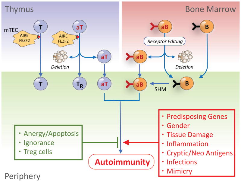
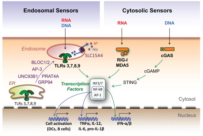
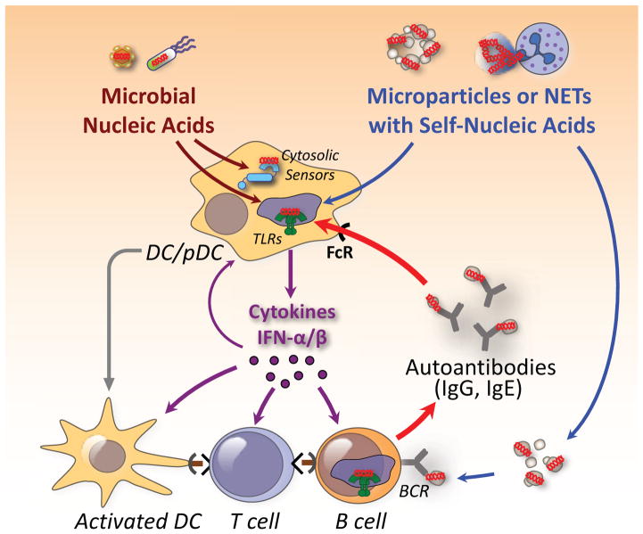
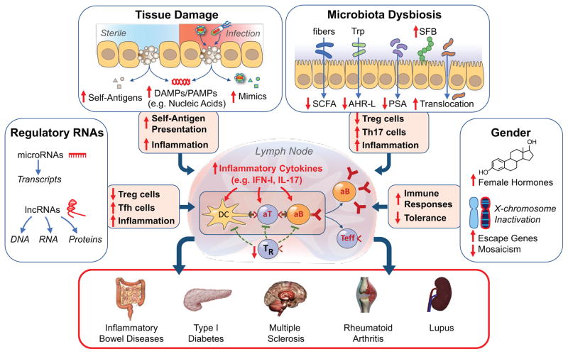
Efforts to understand autoimmunity have been pursued relentlessly for several decades. It has become apparent that the immune system evolved multiple mechanisms for controlling self-reactivity, and defects in one or more of these mechanisms can lead to a breakdown of tolerance. Among the multitude of lesions associated with disease, the most common seem to affect peripheral tolerance rather than central tolerance. The initial trigger for both systemic autoimmune disorders and organ-specific autoimmune disorders probably involves the recognition of self or foreign molecules, especially nucleic acids, by innate sensors. Such recognition, in turn, triggers inflammatory responses and the engagement of previously quiescent autoreactive T cells and B cells. Here we summarize the most prominent autoimmune pathways and identify key issues that require resolution for full understanding of pathogenic autoimmunity.


Conflict of interest statement

Competing Financial Interests

The authors declare no competing financial interests.

Figures

Figure 1
Figure 1. Escape of autoreactive T and B cells from central tolerance and engagement in the periphery
During differentiation, T and B cell precursors with self-reactivity are positively selected in the thymic cortex and bone marrow, respectively, and those with low avidity for self are exported to the periphery. In contrast, autoreactive T cells (aT) with high avidity for self-antigens expressed by medullary thymic epithelial cells (mTEC) under the control of AIRE or FEZF2 are deleted or differentiate to Treg cells (TR), while autoreactive B cells (aB) are deleted or receptor-edited. Central tolerance, however, is incomplete, and some autoreactive T and B cells are exported to the periphery. The exported cells are normally controlled by peripheral tolerance mechanisms, including inhibitory molecules, anergy, ignorance and suppression by Treg cells. However, in genetically-predisposed individuals, tissue damage, inflammation, and presentation of sequestered, cryptic, neo self-antigens or microbial mimics might provoke break of tolerance and autoimmunity.
Figure 2
Figure 2. Engagement of endosomal or cytosolic nucleic acid sensors as central events in inflammatory responses
Nucleic acid sensors are critical innate immune receptors that reside either in endolysosomes or the cytosol. Upon recognition of specific ligands, they initiate a signaling cascade resulting in the activation of several transcription factors that promote cell activation and production of type I interferons (IFN-I) and inflammatory cytokines. Two ER molecules, GRP94 and PRAT4A, act in concert to ensure proper folding of TLRs 3, 7, 8 and 9 and exit from the ER, while UNC93B1 mediates TLR transport to endolysosomes, where ligand recognition takes place. Other proteins participating in TLR trafficking and/or function are the adaptor protein 3 (AP-3), the biogenesis of lysosome organelle complex 1 and 2 (BLOC1/2), and the solute carrier 15A4 (SLC15A4), a molecule known to transport protons (H+), histidine (His) and selected peptides from endosomes to the cytosol. In the cytosol, RNA is sensed by the helicases RIG-I and MDA5, while DNA is primarily sensed by the cyclic GMP-AMP synthase (cGAS). Engagement of cGAS leads to synthesis of the second messenger cGAMP that interacts with the stimulator of interferon genes (STING) to promote inflammatory responses.
Figure 3
Figure 3. Pathways by which self and foreign nucleic acid sensors promote autoimmunity
It is postulated that self-nucleic acids in microparticles released from dying cells or in neutrophil extracellular traps (NETs) gain access to acidified endolysosomal compartments of pDCs, DCs, and antigen-specific B cells. TLR engagement and production of inflammatory cytokines causes upregulation of MHC and costimulatory molecules in these cells, antigen presentation, and engagement of autoreactive T cells. Complexes of autoantibodies (IgG, IgE) with nucleic acid-associated molecules are taken up through the FcR and amplify and sustain the inflammatory response. In certain instances, microbial nucleic acids alone or in conjunction with self-nucleic acids released from damaged tissues may constitute the initial trigger.
Figure 4
Figure 4. The multiple pathways to autoimmunity
We posit that autoimmunity may result from disturbances in multiple processes acting singly or in combination. Tissue damage under sterile conditions or due to infections may lead to availability of nucleic acids and other damage- or pathogen-associated molecular patterns (DAMPs, PAMPs), presentation of self-antigens to non-tolerant lymphocytes, and induction of inflammatory responses. Microbiota dysbiosis may result in displacement of beneficial commensals, reductions of several anti-inflammatory factors (short chain fatty acids, SCFA; Aryl hydrocarbon receptor ligands, AHR-L; polysaccharide A, PSA), expansion of adherent bacteria (e.g. segmented filamentous bacteria, SFB in mice), damage of the mucosal/epithelial barrier, and translocation of bacteria and inflammatory products to mesenteric lymph nodes. These effects lead to engagement of TLRs and other innate sensors, production of inflammatory cytokines, reduction in Treg cells (TR), expansion of TH17 and other effector cells, and production of autoantibodies, resulting in organ-specific or systemic autoimmune diseases. Additional autoimmunity-contributing factors may include abnormalities in non-coding regulatory RNAs, gender-associated hormonal effects, and incomplete X chromosome inactivation. These processes require a predisposing genetic background for the pathogenic phenotype to be expressed.

Similar articles

Cited by

References

    1. Kono DH, Theofilopoulos AN. Kelley and Firestein’s Textbook of Rheumatology. 10. Elsevier; Philadelphia: 2017. Autoimmunity; pp. 301–317.
    1. Park H, Bourla AB, Kastner DL, Colbert RA, Siegel RM. Lighting the fires within: the cell biology of autoinflammatory diseases. Nat Rev Immunol. 2012;12:570–580. - PMC - PubMed
    1. Gutierrez-Arcelus M, Rich SS, Raychaudhuri S. Autoimmune diseases - connecting risk alleles with molecular traits of the immune system. Nat Rev Genet. 2016;17:160–174. - PMC - PubMed
    1. Hunt KA, et al. Negligible impact of rare autoimmune-locus coding-region variants on missing heritability. Nature. 2013;498:232–235. - PMC - PubMed
    1. Bouneaud C, Kourilsky P, Bousso P. Impact of negative selection on the T cell repertoire reactive to a self-peptide: a large fraction of T cell clones escapes clonal deletion. Immunity. 2000;13:829–840. - PubMed