. 2017 Aug 8;117(4):525-534.
doi: 10.1038/bjc.2017.181. Epub 2017 Jun 20.

MiR-646 inhibited cell proliferation and EMT-induced metastasis by targeting FOXK1 in gastric cancer

Affiliations

MiR-646 inhibited cell proliferation and EMT-induced metastasis by targeting FOXK1 in gastric cancer

P Zhang et al. Br J Cancer. .

Abstract

Background: MiR-646 has been reported to be aberrantly expressed in human cancers. However, the underlying molecular mechanisms of action of miR-646 in gastric cancer (GC) have not yet been investigated.

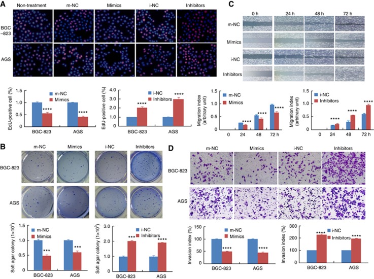
Methods: In vitro function of miR-646 in GC was evaluated using EdU assay, plate colony formation assay, and matrigel invasion assay. Real-time PCR or western blotting was performed to detect miR-646 and FOXK1 expressions. In vivo tumour growth and metastasis were conducted in nude mice.

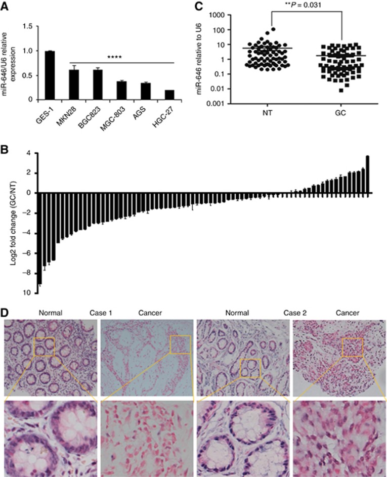
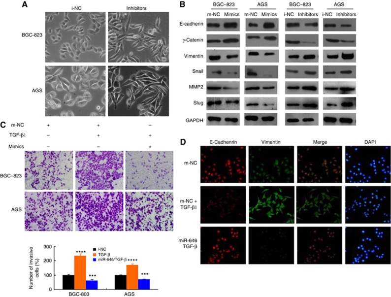
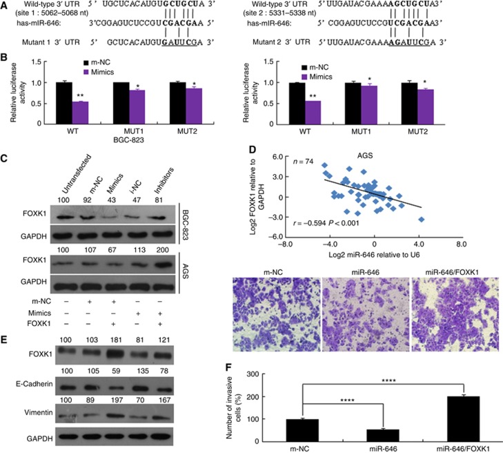
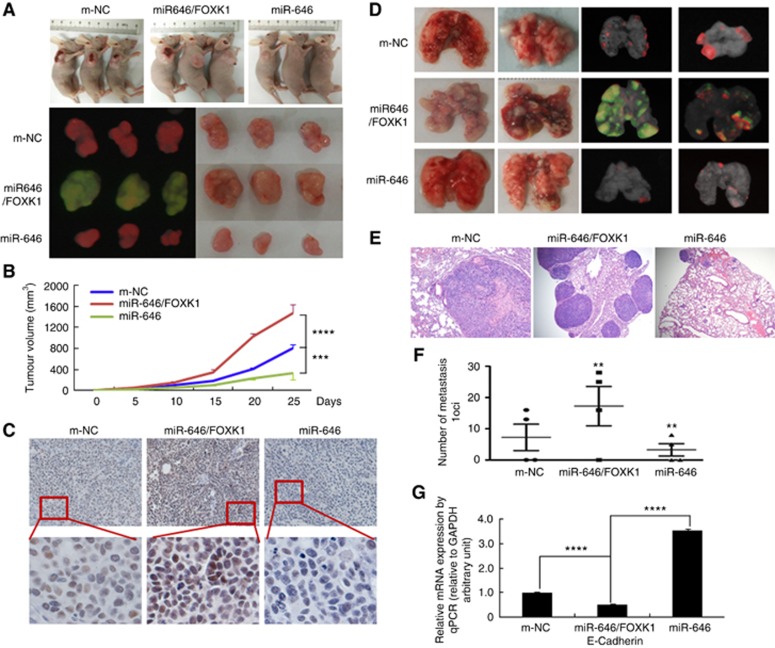
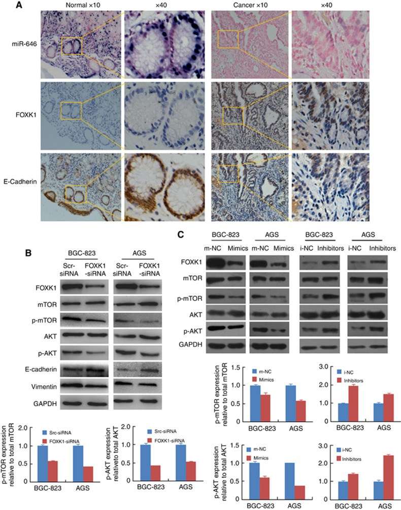
Results: MiR-646 expression was downregulated in GC tissues compared with adjacent normal tissues. Low miR-646 expression is associated with malignant progression. Transient transfection of GC cells with miR-646 inhibited their growth and migration. Moreover, miR-646 influenced the expression of epithelial-mesenchymal transition (EMT)-associated proteins. TGF-β1 treatment significantly suppressed the expression of miR-646 and overexpression of this microRNA counteracted the influence of the TGF-β1-induced EMT phenotype. In terms of the underlying mechanism, miR-646 directly targeted FOXK1. In vivo, it inhibited the FOXK1-mediated proliferation and EMT-induced metastasis. Consistently, inverse correlations were also observed between the expression of miR-646 and FOXK1 in human GC tissue samples. Furthermore, miR-646 regulated Akt/mTOR signalling after FOXK1.

Conclusions: miR-646 inhibited GC cell proliferation and the EMT progression in GC cells by targeting FOXK1.


Conflict of interest statement

The authors declare no conflict of interest.

Figures

Figure 1
Figure 1
miR-646 expression is downregulated in GC cell lines and tissues. (A) Decreased miR-646 expression was detected in all five GC cell lines compared with the normal human gastric epithelial cell line GES-1. ****P<0.001. (B) Real-time PCR analysis of miR-646 expression in 74 pairs of human GC tissues and their adjacent normal mucosal tissues. The error bars represent the mean±s.d. from three independent experiments. (C) As analysed by qRT–PCR, miR-646 expression level in GC tissues was significantly lower than that observed in the corresponding noncancerous gastric mucosa tissue. **P<0.05 (D) Representative ISH images of miR-646 expression. Scale bars, 50 μm.
Figure 2
Figure 2
Functional analysis of miR-646 in vitro. (A) DNA synthesis in GC cells was measured by EdU incorporation assay at 48 h after the indicated transfection. Red fluorescence represents the EdU-positive cells; blue fluorescence from the Hoechst stain represents the total cells. ****P<0.001, mimics vs m-NC; ****P<0.001, inhibitor vs i-NC. (B) Effects of miR-646 mimics or inhibitor on the proliferation of GC cell lines, as determined by anchorage-independent growth ability assay. ***P<0.01. (C) Cell migration activity was measured by wound healing assay at 48 h after transfection with miR-646. ****P<0.001 and ***P<0.01. (D) To evaluate cell invasiveness, transwell assays were performed on miR-646. Cells were counted under a microscope in five randomly selected fields. ****P<0.001. Scale bars, 100 μm in A.
Figure 3
Figure 3
miR-646 inhibits the EMT in TGF-β-treated GC cells. (A) Morphology of the miR-646 expression levels with i-NC and inhibitor cells, as visualised by phase-contrast microscopy. (B) The expression of EMT biomarkers, including E-cadherin, s-catenin, vimentin, Snail, MMP2, and Slug, was detected by western blotting at 48 h after transfection. (C) Representative figures and data from transwell assays performed using GC cells treated with miR-646 mimics and/or TGF-β (5 ng ml−1) for 24 h. ***P<0.01 and ****P<0.001. (D) Immunofluorescence staining for the expression of E-cadherin (red) and vimentin (green) in BCG-823 cells treated with miR-646 mimics and TGF-β (5 ng ml−1), TGF-β only or m-NC. DAPI was used to show the locations of nuclei (blue). Representative immunofluorescence images were captured. The scale bars represent 20 μm in A and D.
Figure 4
Figure 4
FOXK1 is a direct target of miR-646. (A) The putative miR-646-binding sites in the FOXK1 3′-UTR; nt, nucleotides. (B) Luciferase activity in the indicated GC cells following transfection of conserved miR-646 binding site-mutated 3′-UTR-driven reporter constructs; wt, wild type; mut, mutant; site 1, the miR-646 motif spanning nt 5062–5068 nt; site 2, the motif spanning nt 5331–5338 nt in the FOXK1 3′-UTR; n=3. **P<0.05 and *P>0.05. (C) Western blot analysis of FOXK1 protein expression in GC cells transfected with miR-646 mimics or inhibitor. Untransfected cells, as well as cells transfected with a control mimic or inhibitor plasmid, were used as controls. (D) In human GC tissues, FOXK1 was negatively correlated with miR-646 at the mRNA level (n=74). (E) The FOXK1, E-cadherin, and vimentin protein levels were detected by western blotting in cells transfected with a FOXK1 expression plasmid, miR-646 mimics, or m-NC. The relative protein expression levels were quantified by comparing the gray level of each band using Quantity One Software (Life Science Research, Hercules, CA, USA). (F) Migratory activity of cells transfected with a FOXK1 expression plasmid and/or miR-646 mimics was evaluated by transwell invasion assay. ****P<0.001. All of these experiments were repeated three times with identical findings.
Figure 5
Figure 5
miR-646 regulated GC tumour growth and metastasis by targeting FOXK1 in vivo. (A) Fluorescence images of subcutaneous tumours from mice injected with BGC-823/m-NC, BGC-823/miR-646/FOXK1, or BGC-823/miR-646 cells. (B) Tumour size was measured at 5 days after tumour cell inoculation in each group. ***P<0.01, m-NC vs miR-646/FOXK1; and ****P<0.001, miR-646 vs miR-646/FOXK1. (C) Immunohistochemical (IHC) staining of FOXK1 expression in subcutaneous tumours from mice injected with BGC-823/m-NC, BGC-823/miR-646/FOXK1, or BGC-823/miR-646 cells. (D) Mice were orthotopically transplanted with BGC-823 cells (n=3 in each group). (E) Metastatic cancer tissues were stained with H&E. (F) The number of metastatic loci in the lungs was counted. **P<0.05, m-NC vs miR-646/FOXK1 and miR-646/FOXK1 vs miR-646. (G) The expression of E-cadherin in tumours derived from BGC-823 cells was determined by quantitative PCR. ****P<0.001, m-NC vs miR-646/FOXK1; miR-646 vs miR-646/FOXK1. Scale bars, 200 μm in C and 400 μm in E.
Figure 6
Figure 6
miR-646 suppresses the EMT via Akt/mTOR signalling. (A) ISH analysis of miR-646 and IHC analysis of FOXK1 and E-cadherin were performed. These figures were the representatives of gastric tissues from 10 cancerous and 10 non-cancerous patients. The scale bars represent 200 μm in A. (B) Knockdown of FOXK1 by siRNA in gastric cancer cell decreased the protein expression of E-cadherin, vimentin and phosphorylation of Akt and mTOR. (C) FOXK1, mTOR, p-mTOR, Akt, and p-Akt protein expression in BGC-823 and AGS cells transfected with the miR-646 mimic, inhibitor, or corresponding control (NC) plasmid.

References

    1. Calin GA, Croce CM (2006) MicroRNA-cancer connection: the beginning of a new tale. Cancer Res 66: 7390–7394. - PubMed
    1. Chang L, Graham PH, Hao J, Ni J, Bucci J, Cozzi PJ, Kearsley JH, Li Y (2013) Acquisition of epithelial-mesenchymal transition and cancer stem cell phenotypes is associated with activation of the PI3K/Akt/mTOR pathway in prostate cancer radioresistance. Cell Death Dis 4: e875. - PMC - PubMed
    1. Dong P, Ihira K, Xiong Y, Watari H, Hanley SJ, Yamada T, Hosaka M, Kudo M, Yue J, Sakuragi N (2016) Reactivation of epigenetically silenced miR-124 reverses the epithelial-to-mesenchymal transition and inhibits invasion in endometrial cancer cells via the direct repression of IQGAP1 expression. Oncotarget 7: 20260–20270. - PMC - PubMed
    1. Dwivedi SK, Mustafi SB, Mangala LS, Jiang D, Pradeep S, Rodriguez-Aguayo C, Ling H, Ivan C, Mukherjee P, Calin GA, Lopez-Berestein G, Sood AK, Bhattacharya R (2016) Therapeutic evaluation of microRNA-15a and microRNA-16 in ovarian cancer. Oncotarget 7: 15093–15104. - PMC - PubMed
    1. Fu W, Tao T, Qi M, Wang L, Hu J, Li X, Xing N, Du R, Han B (2016) MicroRNA-132/212 upregulation inhibits TGF-β-mediated epithelial-mesenchymal transition of prostate cancer cells by targeting SOX4. Prostate 76: 1560–1570. - PubMed

MeSH terms