

Prevalence and orthopedic management of foot and ankle deformities in Charcot-Marie-Tooth disease
- PMID: 28632967
- PMCID: PMC5811923
- DOI: 10.1002/mus.25724
Prevalence and orthopedic management of foot and ankle deformities in Charcot-Marie-Tooth disease
Abstract
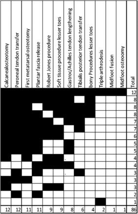
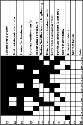
Introduction: Foot deformities are frequent complications in Charcot-Marie-Tooth disease (CMT) patients, often requiring orthopedic surgery. However, there are no prospective, randomized studies on surgical management, and there is variation in the approaches among centers both within and between countries.
Methods: In this study we assessed the frequency of foot deformities and surgery among patients recruited into the Inherited Neuropathies Consortium (INC). We also designed a survey addressed to orthopedic surgeons at INC centers to determine whether surgical approaches to orthopedic complications in CMT are variable.
Results: Foot deformities were reported in 71% of CMT patients; 30% of the patients had surgery. Survey questions were answered by 16 surgeons working in different specialized centers. Most of the respondents were foot and ankle surgeons. There was marked variation in surgical management.
Discussion: Our findings confirm that the approaches to orthopedic management of CMT are varied. We identify areas that require further research. Muscle Nerve 57: 255-259, 2018.
Keywords: Charcot-Marie-Tooth disease; foot deformities; foot surgery; orthopedic complications; pes cavus; survey.
© 2017 The Authors Muscle & Nerve Published by Wiley Periodicals, Inc.
Figures
References
-
- Reilly MM, Murphy SM, Laura M. Charcot‐Marie‐Tooth disease. J Periph Nerv Syst 2011;16:1–14. - PubMed
-
- Alexander JF, Johnson KA. Assessment and management of pes cavus in Charcot‐Marie‐Tooth disease. Clin Orthop Relat Res 1989;246:273–281. - PubMed
-
- Holmes JR, Hansen ST Jr. Foot and ankle manifestations of Charcot‐Marie‐Tooth disease. Foot Ankle 1993;14:476–486. - PubMed
-
- Faldini C, Traina F, Nanni M, Mazzotti A, Calamelli C, Fabbri D, et al.. Surgical treatment of cavus foot in Charcot‐Marie‐tooth disease: a review of twenty‐four cases: AAOS exhibit selection. J Bone Joint Surg Am 2015;97:e30. - PubMed
Publication types
MeSH terms
Grants and funding
LinkOut - more resources
Full Text Sources
Other Literature Sources
Medical
