. 2017 Jun 19;41(6):674-684.e5.
doi: 10.1016/j.devcel.2017.05.014.

Apoptosis and Compensatory Proliferation Signaling Are Coupled by CrkI-Containing Microvesicles

Affiliations

Apoptosis and Compensatory Proliferation Signaling Are Coupled by CrkI-Containing Microvesicles

Kajal H Gupta et al. Dev Cell. .

Abstract

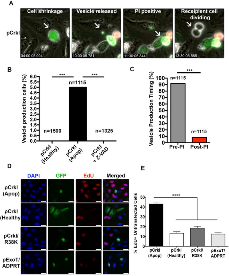
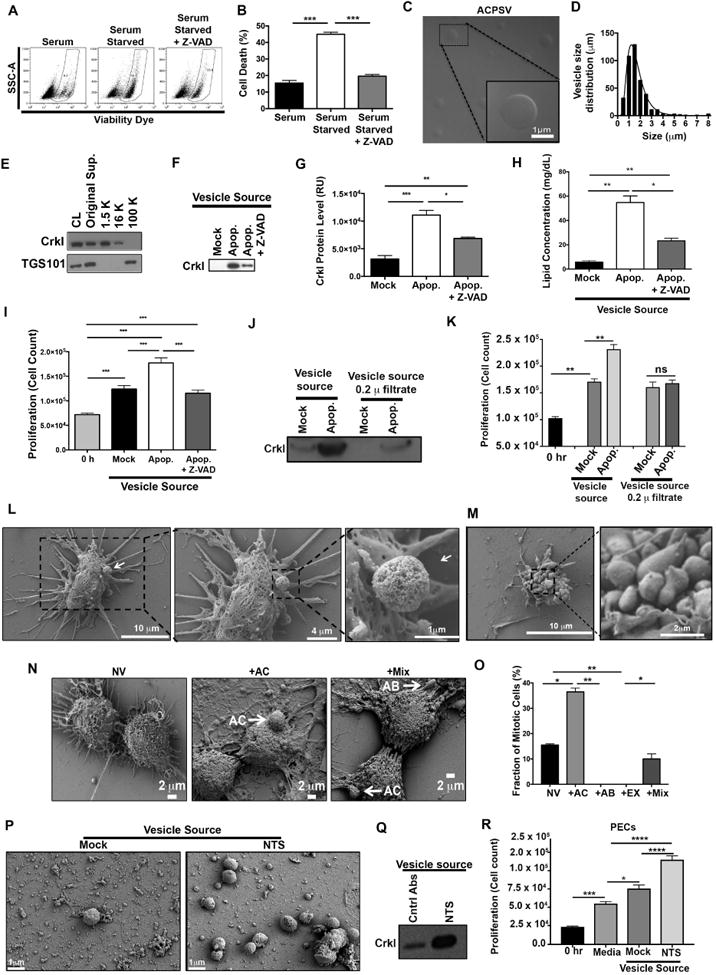
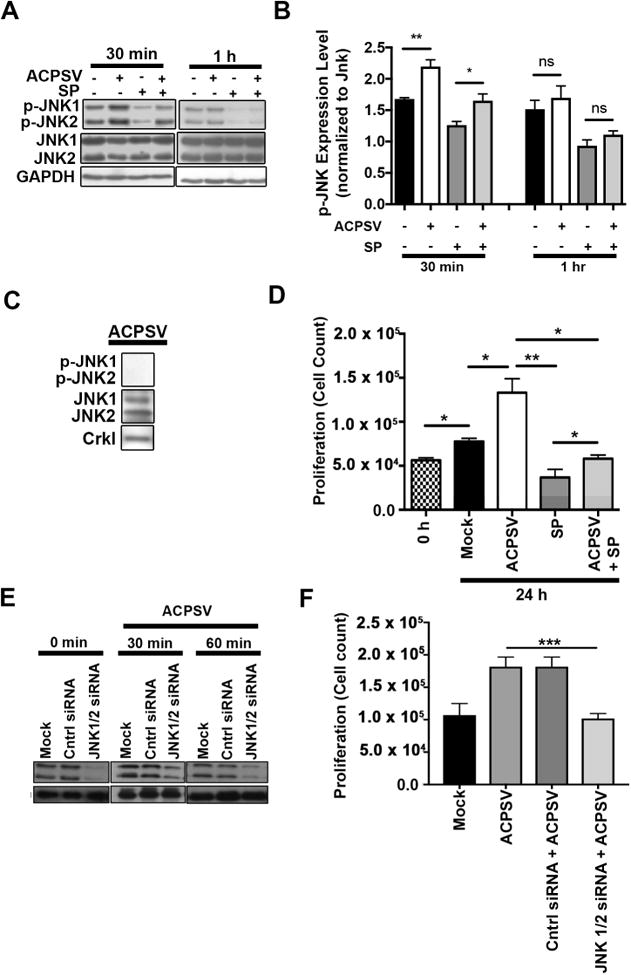
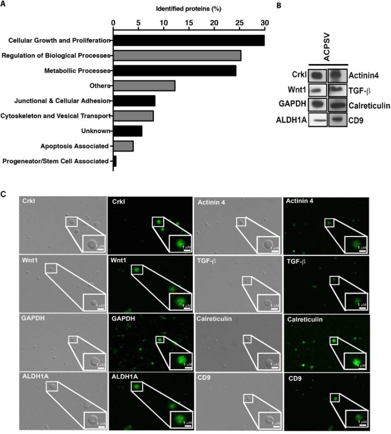
Apoptosis has been implicated in compensatory proliferation signaling (CPS), whereby dying cells induce proliferation in neighboring cells as a means to restore homeostasis. The nature of signaling between apoptotic cells and their neighboring cells remains largely unknown. Here we show that a fraction of apoptotic cells produce and release CrkI-containing microvesicles (distinct from exosomes and apoptotic bodies), which induce proliferation in neighboring cells upon contact. We provide visual evidence of CPS by videomicroscopy. We show that purified vesicles in vitro and in vivo are sufficient to stimulate proliferation in other cells. Our data demonstrate that CrkI inactivation by ExoT bacterial toxin or by mutagenesis blocks vesicle formation in apoptotic cells and inhibits CPS, thus uncoupling apoptosis from CPS. We further show that c-Jun amino-terminal kinase (JNK) plays a pivotal role in mediating vesicle-induced CPS in recipient cells. CPS could have important ramifications in diseases that involve apoptotic cell death.

Keywords: ACPS; ACPSV; CT10 regulator of kinase I; CrkI; ExoT; Exotoxin T; JNK; NTS; Pseudomonas aeruginosa; apoptosis; apoptotic compensatory proliferation signaling; apoptotic compensatory proliferation signaling vesicle; c-Jun amino-terminal kinase; glomerulonephritis; nephritis; nephrotoxic serum.


Conflict of interest statement

Conflict of Interest: A US divisional patent has been filed by Rush University Medical Center, based on research data included in this manuscript.

Figures

Fig. 1
Fig. 1. Apoptotic cells produce and release CrkI-containing vesicles, which appear to be capable of inducing proliferation in bystander cells
HeLa cells were transfected with CrkI-GFP in the presence or absence of Z-VAD and followed by IF time-lapse videomicroscopy. (A) Selected movie frames of a CrkI-transfected apoptotic cell are shown. This cell releases a CrkI-containing microvesicle, before it becomes PI positive, and the bystander cell initiates mitosis after contacting this vesicle. (B) The percentages of non-apoptotic, apoptotic, and Z-VAD-treated, CrkI-GFP transfected cells producing vesicle is shown. (C) The ability of CrkI-GFP transfected apoptotic cells to produce and release vesicles prior or post PI uptake was assessed by IF time-lapse microscopy and the tabulated results are shown. (For B & C, n≥1115, *** p<0.001, χ2 analysis). (D-E) Cells were transiently transfected with CrkI-GFP, CrkI/R38K-GFP, or ExoT/ADPRT-GFP expression vectors. 17h after transfection, cells were treated with EdU for 2h, fixed by 10% TCA, and analyzed for EdU incorporation in untransfected cells surrounding transfected apoptotic cells (identified by cell shrinkage/rounding) or healthy (identified by spread-out morphology). Representative images are shown in (D) and the tabulated data are shown in (E) (n≥3 independent experiments, 10 random fields/experiment, **** p<0.0001, One-way ANOVA).
Fig. 2
Fig. 2. Purification and characterization of CrkI-containing microvesicles (ACPSVs)
(A) HeLa cells either received 10% serum (Mock) or were induced to undergo apoptosis by serum-starvation in the presence or absence of Z-VAD. 24h after serum starvation, apoptotic cell death was assessed by flow cytometry and the tabulated results from 3 independent experiments are shown in (B). (*** p<0.001, One-way ANOVA). (C-I) Supernatants from the indicated cultures were fractionated by differential centrifugation and evaluated for their CrkI-containing microvesicles (ACPSVs) and CPS. (C) ACPSVs in the 16K fraction of apoptotic cells were visualized by DIC. (D) Vesicle size distribution was determined and plotted (Mean size ± S.D. = 1.76 ± 1.04 μm, n=822). (E) Peptides from the cell lysate (CL) and the indicated fractions of serum-starved apoptotic cells were probed for TGS101 (exosome marker) and CrkI (ACPSV marker) by Western blotting. (F) The 16K fractions of healthy (Mock), serum-starved (Apop.) and Z-VAD treated serum-starved (Apop. + Z-VAD) were probed for ACPSV content by Western blotting of CrkI and the results from 6 independent studies were plotted in (G). (* p<0.05, ** p<0.01, *** p<0.001, One-way ANOVA). (H) The lipid contents of the vesicles in the 16K fractions of the indicated cultures were measured and the tabulated data from 3 experiments are shown. (* p<0.01, ** p<0.005, One-way ANOVA). (I) HeLa cells were treated with 16K fractions of serum-fed healthy (Mock), serum-starved apoptotic (Apop.), or Z-VAD treated serum-starved (Apop. + Z-VAD) and the proliferation levels were determined by cell count 24h after treatment. The tabulated data from n≥6 independent experiments are shown as the Mean ± S.D. (*** p<0.001, One-way ANOVA). (J-K) The 16K fractions of mock and apoptotic (apop.) HeLa cells were passaged through 0.2 μ filter to remove vesicle. (L-M) HeLa cells were seeded on a coverslip and induced to undergo apoptosis by serum-starvation. 24h after serum starvation, cells were fixed and analyzed by SEM. (L) Representative images of a vesicle producing cell are shown. (M) Representative images of an apoptotic body-producing cell are shown. (N-O) Original supernatant from serum-starved apoptotic HeLa cell culture was spun down at 100,000× g (100K) to collect all vesicles, which were then added to adherent HeLa cells on a coverslip. Five hours after vesicle addition, the fractions of mitotic cells, with no vesicle (NV); with ACPSV only (+AC); with apoptotic body only (+AB); or with a combination of vesicles (+Mix) were assessed by SEM. Selected images representing each group is shown in (N) and the tabulated data from 3 experiments are shown in (O). (** p ≤ 0.01, *** p ≤ 0.001, **** p ≤ 0.0001; One-way ANOVA). (P-Q) C57BL/6 mice were injected with nephrotoxic serum (NTS) to induce nephritis or isotype antibody control (Mock). Vesicles were harvested from mock and NTS-injected mice glomeruli and imaged by SEM (P) and evaluated for their CrkI contents by Western blot (Q), and for their ability to induce proliferation in adherent kidney parietal epithelial cells (PECs) in (R). (3 independent experiment, 4 mice/group, each group was done in triplicates, * p<0.05, *** p<0.001, **** p<0.0001, One-way ANOVA).
Fig. 3
Fig. 3. JNK activity is required for ACPSV-induced compensatory proliferation in bystander cells
(A) The impact of ACPSVs on JNK1/2 activation (phosphorylation) in recipient HeLa cells in the presence and absence of 20μM JNK-specific inhibitor, SP600125 (SP), was assessed by Western blotting at 30 min and 1h after treatment with 16K fractions from serum-fed healthy (no ACPSV) or apoptotic (contains ACPSV) HeLa cells. Phospho JNK1/2 (p-JNK1/2) expression levels were normalized to total JNK1/2 protein levels and plotted in (B). Data are shown as Mean ± SEM (n=3, * p ≤ 0.05, ** p ≤ 0.01, One-way ANOVA). (C) ACPSVs were probed for phosphorylated and unphosphorylated JNK1/2. (D) HeLa cells were pre-treated with SP or DMSO (0.1%) for 2h prior to treatment with ACPSV or Mock. Proliferation was determined by measuring total cell counts at the time of treatment (0h) and 24h post treatment. Data are shown as Mean ± SEM (n=3, * p ≤ 0.05, ** p ≤ 0.01, *** p ≤ 0.001, One-way ANOVA). (E-F) HeLa cells were treated with JNK1/2-specific siRNA or scrambled control (Mock), 24h prior to ACPS vesicle treatment. (E) JNK1/2 levels were evaluated by Western blotting in HeLa recipient cells prior to vesicle treatment (0 mins) or 30 or 60 mins after vesicle treatment. (F) The ability of ACPSVs to induce proliferation in control or JNK1/2-depleted HeLa cells was evaluated 24h after vesicle addition by cell counts. The data indicate that JNK function is required in recipient cells to mediate CPS induced by ACPSVs. (n=3, ** p ≤ 0.001).
Fig. 4
Fig. 4. Proteomic analyses of ACPSV by LC- MS/MS
Peptides from the 16K fractions of serum-starved apoptotic HeLa cells were analyzed by LC-MS/MS. (A) Some of the notable protein groupings were plotted based on the number of proteins identified in each grouping. The results of LC-MS/MS analyses were further corroborated by evaluating the presence of representative proteins from the aforementioned categories within ACPSVs by Western blotting (B) and by IF microscopy (C).

References

    1. Akers JC, Gonda D, Kim R, Carter BS, Chen CC. Biogenesis of extracellular vesicles (EV): exosomes, microvesicles, retrovirus-like vesicles, and apoptotic bodies. J Neurooncol. 2013;113:1–11. - PMC - PubMed
    1. Almeida EA, Ilic D, Han Q, Hauck CR, Jin F, Kawakatsu H, Schlaepfer DD, Damsky CH. Matrix survival signaling: from fibronectin via focal adhesion kinase to c-Jun NH(2)-terminal kinase. J Cell Biol. 2000;149:741–754. - PMC - PubMed
    1. Bergantinos C, Corominas M, Serras F. Cell death-induced regeneration in wing imaginal discs requires JNK signalling. Development. 2010;137:1169–1179. - PubMed
    1. Bouchard V, Harnois C, Demers MJ, Thibodeau S, Laquerre V, Gauthier R, Vezina A, Noel D, Fujita N, Tsuruo T, et al. B1 integrin/Fak/Src signaling in intestinal epithelial crypt cell survival: integration of complex regulatory mechanisms. Apoptosis: an international journal on programmed cell death. 2008;13:531–542. - PubMed
    1. Braun F, Bertin-Ciftci J, Gallouet AS, Millour J, Juin P. Serum-nutrient starvation induces cell death mediated by Bax and Puma that is counteracted by p21 and unmasked by Bcl-x(L) inhibition. PloS one. 2011;6:e23577. - PMC - PubMed

Publication types

MeSH terms