

Microsphere-Based Scaffolds in Regenerative Engineering
- PMID: 28633566
- PMCID: PMC11610505
- DOI: 10.1146/annurev-bioeng-071516-044712
Microsphere-Based Scaffolds in Regenerative Engineering
Abstract
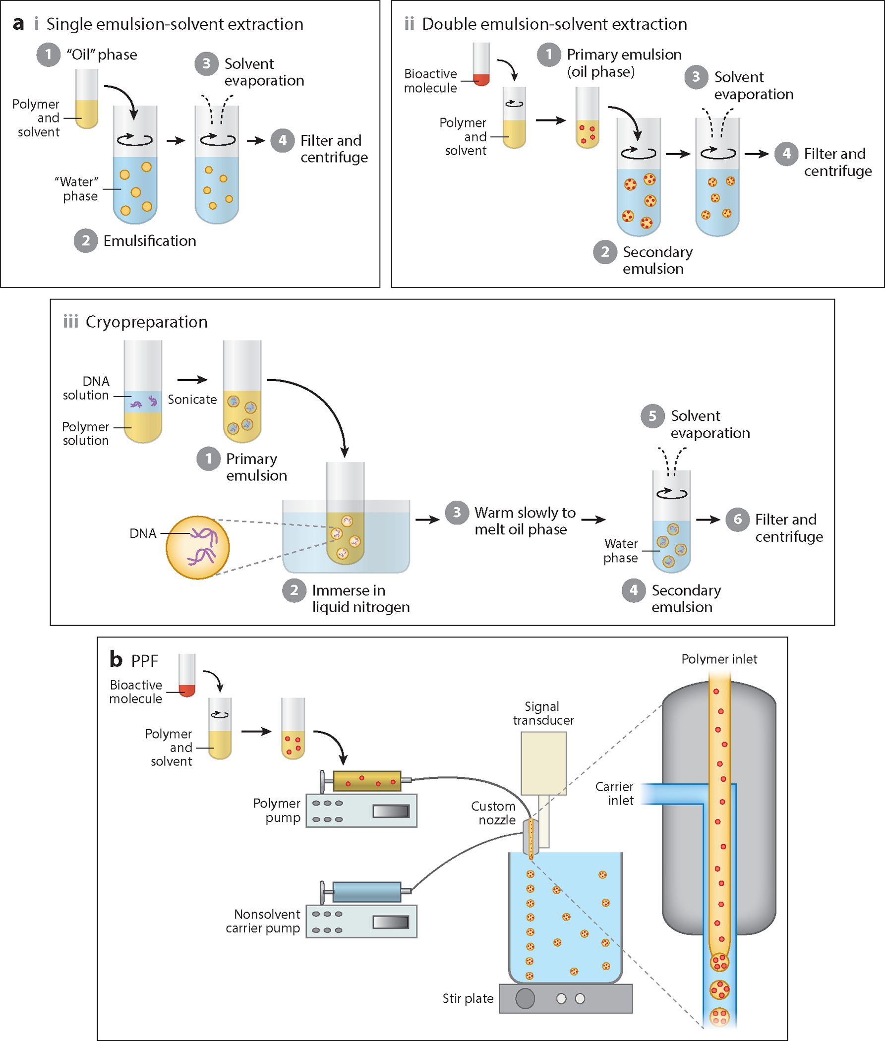
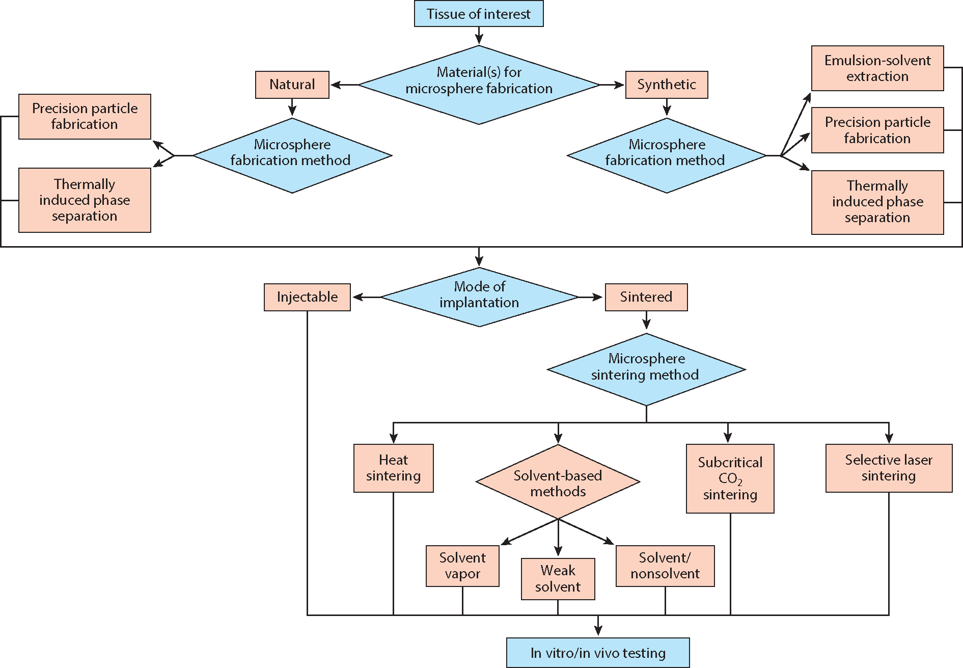
Microspheres have long been used in drug delivery applications because of their controlled release capabilities. They have increasingly served as the fundamental building block for fabricating scaffolds for regenerative engineering because of their ability to provide a porous network, offer high-resolution control over spatial organization, and deliver growth factors/drugs and/or nanophase materials. Because they provide physicochemical gradients via spatiotemporal release of bioactive factors and nanophase ceramics, microspheres are a desirable tool for engineering complex tissues and biological interfaces. In this review we describe various methods for microsphere fabrication and sintering, and elucidate how these methods influence both micro- and macroscopic scaffold properties, with a special focus on the nature of sintering. Furthermore, we review key applications of microsphere-based scaffolds in regenerating various tissues. We hope to inspire researchers to join a growing community of investigators using microspheres as tissue engineering scaffolds so that their full potential in regenerative engineering may be realized.
Keywords: microsphere fabrication; microsphere incorporating scaffolds; microsphere sintering; microsphere-based scaffolds; microspheres.
Conflict of interest statement
DISCLOSURE STATEMENT
C.J.B. is a shareholder and cofounder of Orbis Biosciences. The other authors are not aware of any affiliations, memberships, funding, or financial holdings that might be perceived as affecting the objectivity of this review.
Figures
References
-
- Ravichandran R, Sundarrajan S, Venugopal JR, Mukherjee S, Ramakrishna S. 2012. Advances in polymeric systems for tissue engineering and biomedical applications. Macromol. Biosci. 12:286–311 - PubMed
-
- Subia B, Kundu J, Kundu S. 2010. Biomaterial Scaffold Fabrication Techniques for Potential Tissue Engineering Applications. www.intechopen.com: INTECH
-
- Shi X, Su K, Varshney RR, Wang Y, Wang D-A. 2011. Sintered microsphere scaffolds for controlled release and tissue engineering. Pharm. Res. 28:1224–28 - PubMed
Publication types
MeSH terms
Substances
Grants and funding
LinkOut - more resources
Full Text Sources
Other Literature Sources
